Fluorine »
PDB 5avz-5btd »
5aw1 »
Fluorine in PDB 5aw1: Kinetics By X-Ray Crystallography: Tl+-Substitution of Bound K+ in the E2.MGF42-.2K+ Crystal After 85 Min
Protein crystallography data
The structure of Kinetics By X-Ray Crystallography: Tl+-Substitution of Bound K+ in the E2.MGF42-.2K+ Crystal After 85 Min, PDB code: 5aw1
was solved by
H.Ogawa,
F.Cornelius,
A.Hirata,
C.Toyoshima,
with X-Ray Crystallography technique. A brief refinement statistics is given in the table below:
| Resolution Low / High (Å) | 14.99 / 3.35 |
| Space group | C 1 2 1 |
| Cell size a, b, c (Å), α, β, γ (°) | 222.035, 50.855, 163.971, 90.00, 104.02, 90.00 |
| R / Rfree (%) | 29.7 / 30.5 |
Other elements in 5aw1:
The structure of Kinetics By X-Ray Crystallography: Tl+-Substitution of Bound K+ in the E2.MGF42-.2K+ Crystal After 85 Min also contains other interesting chemical elements:
| Magnesium | (Mg) | 2 atoms |
| Potassium | (K) | 2 atoms |
| Thallium | (Tl) | 3 atoms |
Fluorine Binding Sites:
The binding sites of Fluorine atom in the Kinetics By X-Ray Crystallography: Tl+-Substitution of Bound K+ in the E2.MGF42-.2K+ Crystal After 85 Min
(pdb code 5aw1). This binding sites where shown within
5.0 Angstroms radius around Fluorine atom.
In total 4 binding sites of Fluorine where determined in the Kinetics By X-Ray Crystallography: Tl+-Substitution of Bound K+ in the E2.MGF42-.2K+ Crystal After 85 Min, PDB code: 5aw1:
Jump to Fluorine binding site number: 1; 2; 3; 4;
In total 4 binding sites of Fluorine where determined in the Kinetics By X-Ray Crystallography: Tl+-Substitution of Bound K+ in the E2.MGF42-.2K+ Crystal After 85 Min, PDB code: 5aw1:
Jump to Fluorine binding site number: 1; 2; 3; 4;
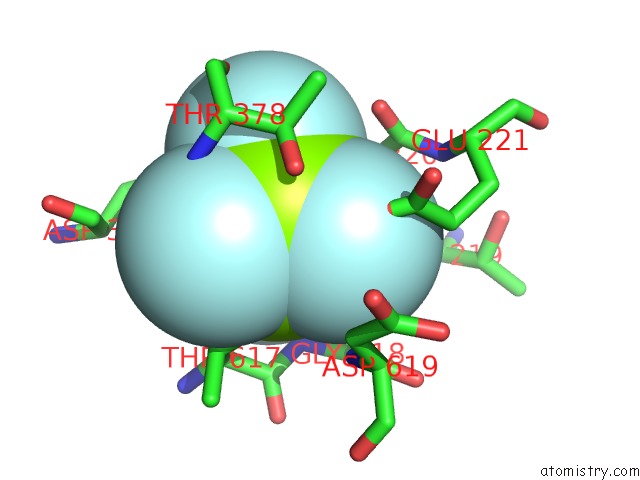
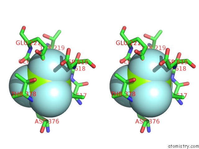
Fluorine binding site 1 out of 4 in 5aw1
Go back to
Fluorine binding site 1 out
of 4 in the Kinetics By X-Ray Crystallography: Tl+-Substitution of Bound K+ in the E2.MGF42-.2K+ Crystal After 85 Min
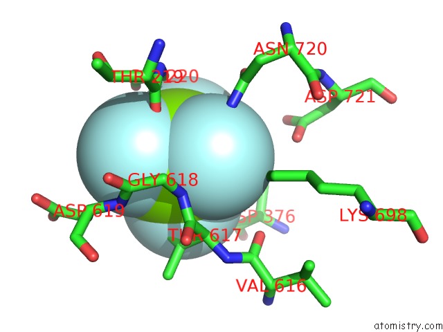
Mono view
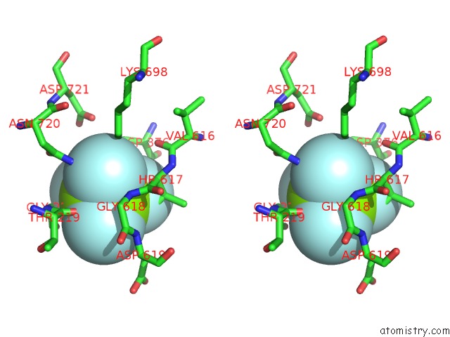
Stereo pair view
Mono view
Stereo pair view
A full contact list of Fluorine with other atoms in the F binding
site number 1 of Kinetics By X-Ray Crystallography: Tl+-Substitution of Bound K+ in the E2.MGF42-.2K+ Crystal After 85 Min within 5.0Å range:
|
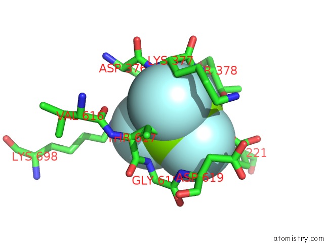
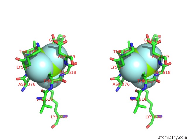
Fluorine binding site 2 out of 4 in 5aw1
Go back to
Fluorine binding site 2 out
of 4 in the Kinetics By X-Ray Crystallography: Tl+-Substitution of Bound K+ in the E2.MGF42-.2K+ Crystal After 85 Min
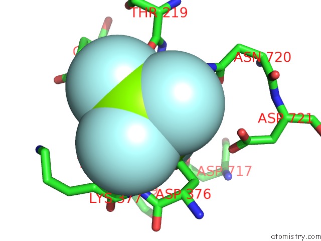
Mono view
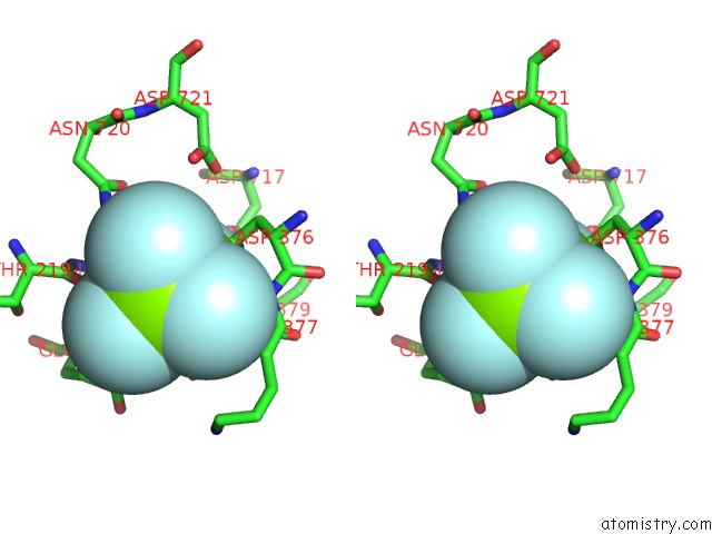
Stereo pair view
Mono view
Stereo pair view
A full contact list of Fluorine with other atoms in the F binding
site number 2 of Kinetics By X-Ray Crystallography: Tl+-Substitution of Bound K+ in the E2.MGF42-.2K+ Crystal After 85 Min within 5.0Å range:
|
Fluorine binding site 3 out of 4 in 5aw1
Go back to
Fluorine binding site 3 out
of 4 in the Kinetics By X-Ray Crystallography: Tl+-Substitution of Bound K+ in the E2.MGF42-.2K+ Crystal After 85 Min
Mono view
Stereo pair view
Mono view
Stereo pair view
A full contact list of Fluorine with other atoms in the F binding
site number 3 of Kinetics By X-Ray Crystallography: Tl+-Substitution of Bound K+ in the E2.MGF42-.2K+ Crystal After 85 Min within 5.0Å range:
|
Fluorine binding site 4 out of 4 in 5aw1
Go back to
Fluorine binding site 4 out
of 4 in the Kinetics By X-Ray Crystallography: Tl+-Substitution of Bound K+ in the E2.MGF42-.2K+ Crystal After 85 Min
Mono view
Stereo pair view
Mono view
Stereo pair view
A full contact list of Fluorine with other atoms in the F binding
site number 4 of Kinetics By X-Ray Crystallography: Tl+-Substitution of Bound K+ in the E2.MGF42-.2K+ Crystal After 85 Min within 5.0Å range:
|
Reference:
H.Ogawa,
F.Cornelius,
A.Hirata,
C.Toyoshima.
Sequential Substitution of K(+) Bound to Na(+),K(+)-Atpase Visualized By X-Ray Crystallography. Nat Commun V. 6 8004 2015.
ISSN: ESSN 2041-1723
PubMed: 26258479
DOI: 10.1038/NCOMMS9004
Page generated: Thu Aug 1 07:57:34 2024
ISSN: ESSN 2041-1723
PubMed: 26258479
DOI: 10.1038/NCOMMS9004
Last articles
F in 4DKPF in 4DKQ
F in 4DK8
F in 4DKO
F in 4DKJ
F in 4DHP
F in 4DBU
F in 4DHM
F in 4DEB
F in 4DC3