Fluorine »
PDB 5ci0-5dde »
5db9 »
Fluorine in PDB 5db9: Structure of Human Dna Polymerase Beta Host-Guest Complex with the N7MG Base Paired with A Dg
Enzymatic activity of Structure of Human Dna Polymerase Beta Host-Guest Complex with the N7MG Base Paired with A Dg
All present enzymatic activity of Structure of Human Dna Polymerase Beta Host-Guest Complex with the N7MG Base Paired with A Dg:
2.7.7.7;
2.7.7.7;
Protein crystallography data
The structure of Structure of Human Dna Polymerase Beta Host-Guest Complex with the N7MG Base Paired with A Dg, PDB code: 5db9
was solved by
M.C.Koag,
S.Lee,
with X-Ray Crystallography technique. A brief refinement statistics is given in the table below:
| Resolution Low / High (Å) | 19.82 / 2.45 |
| Space group | P 1 21 1 |
| Cell size a, b, c (Å), α, β, γ (°) | 54.201, 79.278, 54.720, 90.00, 105.63, 90.00 |
| R / Rfree (%) | 18.7 / 26 |
Other elements in 5db9:
The structure of Structure of Human Dna Polymerase Beta Host-Guest Complex with the N7MG Base Paired with A Dg also contains other interesting chemical elements:
| Sodium | (Na) | 2 atoms |
Fluorine Binding Sites:
The binding sites of Fluorine atom in the Structure of Human Dna Polymerase Beta Host-Guest Complex with the N7MG Base Paired with A Dg
(pdb code 5db9). This binding sites where shown within
5.0 Angstroms radius around Fluorine atom.
In total only one binding site of Fluorine was determined in the Structure of Human Dna Polymerase Beta Host-Guest Complex with the N7MG Base Paired with A Dg, PDB code: 5db9:
In total only one binding site of Fluorine was determined in the Structure of Human Dna Polymerase Beta Host-Guest Complex with the N7MG Base Paired with A Dg, PDB code: 5db9:
Fluorine binding site 1 out of 1 in 5db9
Go back to
Fluorine binding site 1 out
of 1 in the Structure of Human Dna Polymerase Beta Host-Guest Complex with the N7MG Base Paired with A Dg
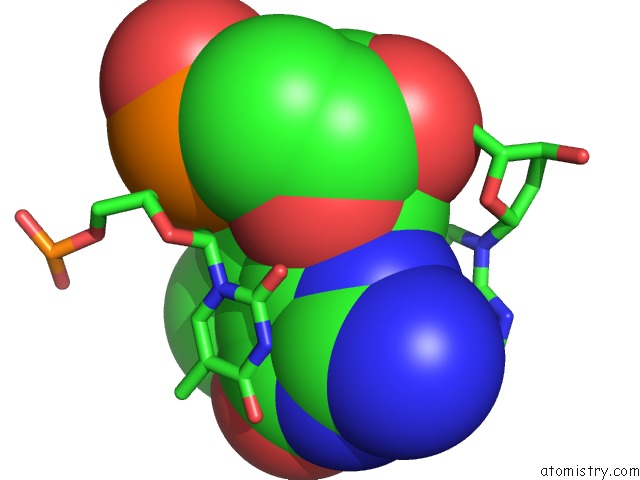
Mono view
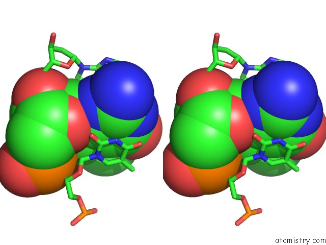
Stereo pair view
Mono view
Stereo pair view
A full contact list of Fluorine with other atoms in the F binding
site number 1 of Structure of Human Dna Polymerase Beta Host-Guest Complex with the N7MG Base Paired with A Dg within 5.0Å range:
|
Reference:
Y.Kou,
M.C.Koag,
S.Lee.
N7 Methylation Alters Hydrogen-Bonding Patterns of Guanine in Duplex Dna. J.Am.Chem.Soc. V. 137 14067 2015.
ISSN: ESSN 1520-5126
PubMed: 26517568
DOI: 10.1021/JACS.5B10172
Page generated: Tue Jul 15 02:59:23 2025
ISSN: ESSN 1520-5126
PubMed: 26517568
DOI: 10.1021/JACS.5B10172
Last articles
Fe in 2YXOFe in 2YRS
Fe in 2YXC
Fe in 2YNM
Fe in 2YVJ
Fe in 2YP1
Fe in 2YU2
Fe in 2YU1
Fe in 2YQB
Fe in 2YOO