Fluorine »
PDB 5ddf-5dy7 »
5duf »
Fluorine in PDB 5duf: Crystal Structure of M. Tuberculosis ECHA6 Bound to Ligand GSK729A
Enzymatic activity of Crystal Structure of M. Tuberculosis ECHA6 Bound to Ligand GSK729A
All present enzymatic activity of Crystal Structure of M. Tuberculosis ECHA6 Bound to Ligand GSK729A:
4.2.1.17;
4.2.1.17;
Protein crystallography data
The structure of Crystal Structure of M. Tuberculosis ECHA6 Bound to Ligand GSK729A, PDB code: 5duf
was solved by
J.A.G.Cox,
G.S.Besra,
K.Futterer,
with X-Ray Crystallography technique. A brief refinement statistics is given in the table below:
| Resolution Low / High (Å) | 51.90 / 1.50 |
| Space group | P 63 |
| Cell size a, b, c (Å), α, β, γ (°) | 103.880, 103.880, 54.390, 90.00, 90.00, 120.00 |
| R / Rfree (%) | 17.3 / 18.4 |
Fluorine Binding Sites:
The binding sites of Fluorine atom in the Crystal Structure of M. Tuberculosis ECHA6 Bound to Ligand GSK729A
(pdb code 5duf). This binding sites where shown within
5.0 Angstroms radius around Fluorine atom.
In total 3 binding sites of Fluorine where determined in the Crystal Structure of M. Tuberculosis ECHA6 Bound to Ligand GSK729A, PDB code: 5duf:
Jump to Fluorine binding site number: 1; 2; 3;
In total 3 binding sites of Fluorine where determined in the Crystal Structure of M. Tuberculosis ECHA6 Bound to Ligand GSK729A, PDB code: 5duf:
Jump to Fluorine binding site number: 1; 2; 3;
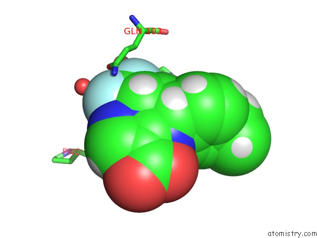
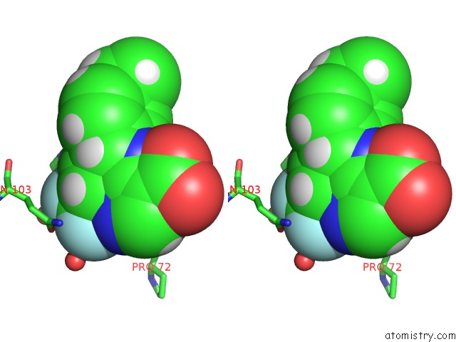
Fluorine binding site 1 out of 3 in 5duf
Go back to
Fluorine binding site 1 out
of 3 in the Crystal Structure of M. Tuberculosis ECHA6 Bound to Ligand GSK729A
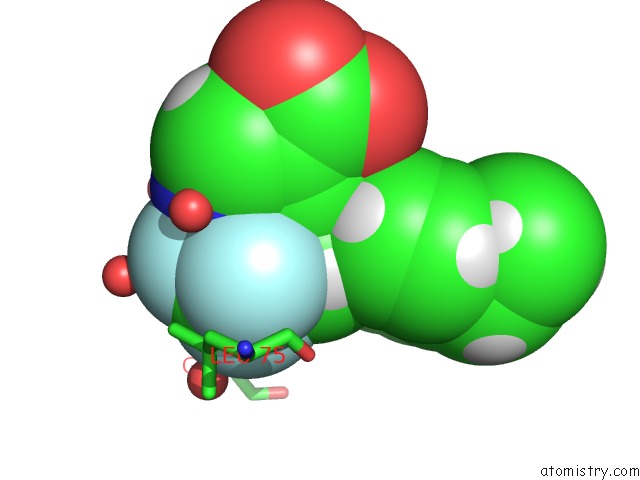
Mono view
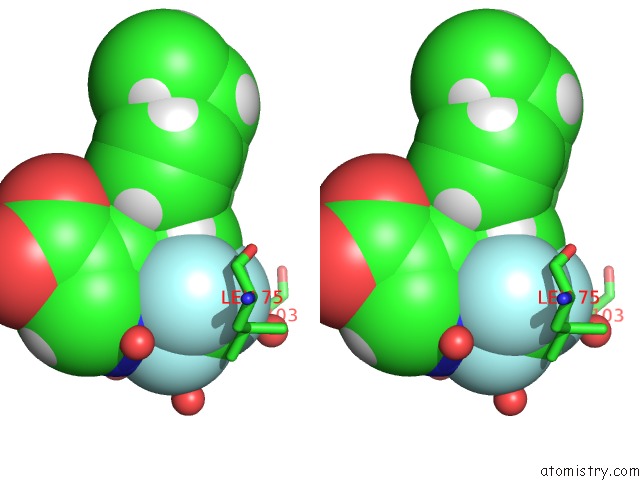
Stereo pair view
Mono view
Stereo pair view
A full contact list of Fluorine with other atoms in the F binding
site number 1 of Crystal Structure of M. Tuberculosis ECHA6 Bound to Ligand GSK729A within 5.0Å range:
|
Fluorine binding site 2 out of 3 in 5duf
Go back to
Fluorine binding site 2 out
of 3 in the Crystal Structure of M. Tuberculosis ECHA6 Bound to Ligand GSK729A
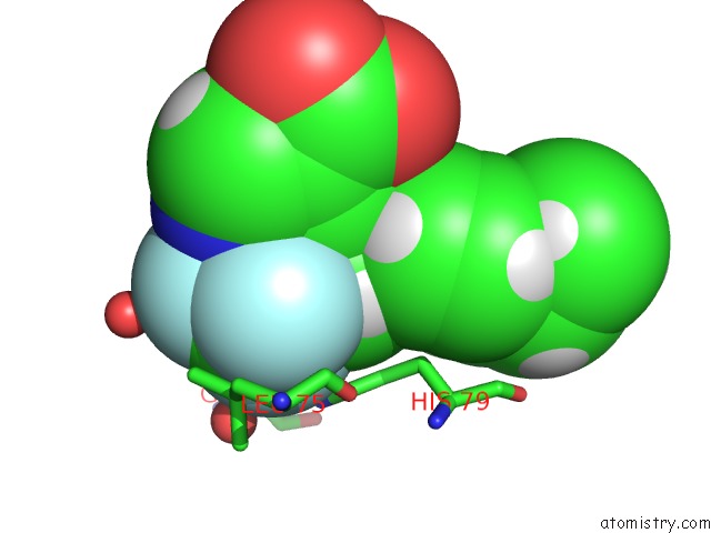
Mono view
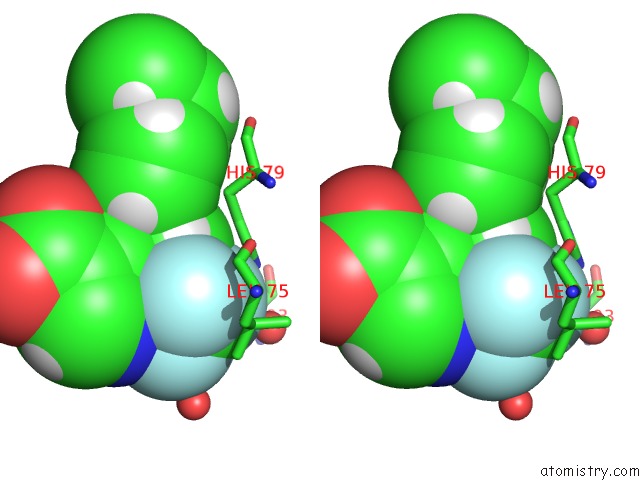
Stereo pair view
Mono view
Stereo pair view
A full contact list of Fluorine with other atoms in the F binding
site number 2 of Crystal Structure of M. Tuberculosis ECHA6 Bound to Ligand GSK729A within 5.0Å range:
|
Fluorine binding site 3 out of 3 in 5duf
Go back to
Fluorine binding site 3 out
of 3 in the Crystal Structure of M. Tuberculosis ECHA6 Bound to Ligand GSK729A
Mono view
Stereo pair view
Mono view
Stereo pair view
A full contact list of Fluorine with other atoms in the F binding
site number 3 of Crystal Structure of M. Tuberculosis ECHA6 Bound to Ligand GSK729A within 5.0Å range:
|
Reference:
J.A.Cox,
K.A.Abrahams,
C.Alemparte,
S.Ghidelli-Disse,
J.Rullas,
I.Angulo-Barturen,
A.Singh,
S.S.Gurcha,
V.Nataraj,
S.Bethell,
M.J.Remuinan,
L.Encinas,
P.J.Jervis,
N.C.Cammack,
A.Bhatt,
U.Kruse,
M.Bantscheff,
K.Futterer,
D.Barros,
L.Ballell,
G.Drewes,
G.S.Besra.
Thpp Target Assignment Reveals ECHA6 As An Essential Fatty Acid Shuttle in Mycobacteria. Nat Microbiol V. 1 15006 2016.
ISSN: ESSN 2058-5276
PubMed: 27571973
DOI: 10.1038/NMICROBIOL.2015.6
Page generated: Thu Aug 1 08:53:42 2024
ISSN: ESSN 2058-5276
PubMed: 27571973
DOI: 10.1038/NMICROBIOL.2015.6
Last articles
Zn in 9J0NZn in 9J0O
Zn in 9J0P
Zn in 9FJX
Zn in 9EKB
Zn in 9C0F
Zn in 9CAH
Zn in 9CH0
Zn in 9CH3
Zn in 9CH1