Fluorine »
PDB 5he2-5hti »
5hf7 »
Fluorine in PDB 5hf7: Tdg Enzyme-Substrate Complex
Enzymatic activity of Tdg Enzyme-Substrate Complex
All present enzymatic activity of Tdg Enzyme-Substrate Complex:
3.2.2.29;
3.2.2.29;
Protein crystallography data
The structure of Tdg Enzyme-Substrate Complex, PDB code: 5hf7
was solved by
E.Pozharski,
S.S.Malik,
A.C.Drohat,
with X-Ray Crystallography technique. A brief refinement statistics is given in the table below:
| Resolution Low / High (Å) | 32.51 / 1.54 |
| Space group | C 1 2 1 |
| Cell size a, b, c (Å), α, β, γ (°) | 97.030, 52.750, 81.340, 90.00, 95.18, 90.00 |
| R / Rfree (%) | 18.6 / 21.4 |
Fluorine Binding Sites:
The binding sites of Fluorine atom in the Tdg Enzyme-Substrate Complex
(pdb code 5hf7). This binding sites where shown within
5.0 Angstroms radius around Fluorine atom.
In total only one binding site of Fluorine was determined in the Tdg Enzyme-Substrate Complex, PDB code: 5hf7:
In total only one binding site of Fluorine was determined in the Tdg Enzyme-Substrate Complex, PDB code: 5hf7:
Fluorine binding site 1 out of 1 in 5hf7
Go back to
Fluorine binding site 1 out
of 1 in the Tdg Enzyme-Substrate Complex
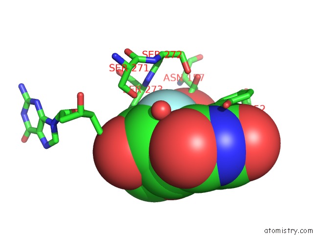
Mono view
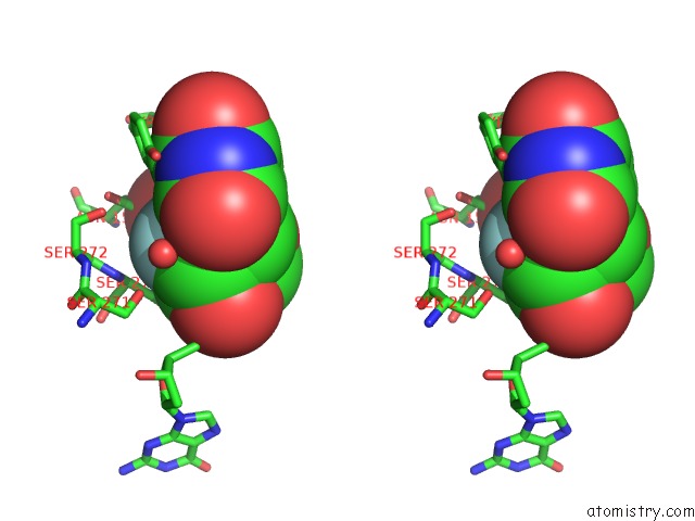
Stereo pair view
Mono view
Stereo pair view
A full contact list of Fluorine with other atoms in the F binding
site number 1 of Tdg Enzyme-Substrate Complex within 5.0Å range:
|
Reference:
C.T.Coey,
S.S.Malik,
L.S.Pidugu,
K.M.Varney,
E.Pozharski,
A.C.Drohat.
Structural Basis of Damage Recognition By Thymine Dna Glycosylase: Key Roles For N-Terminal Residues. Nucleic Acids Res. V. 44 10248 2016.
ISSN: ESSN 1362-4962
PubMed: 27580719
DOI: 10.1093/NAR/GKW768
Page generated: Thu Aug 1 09:50:14 2024
ISSN: ESSN 1362-4962
PubMed: 27580719
DOI: 10.1093/NAR/GKW768
Last articles
Zn in 9J0NZn in 9J0O
Zn in 9J0P
Zn in 9FJX
Zn in 9EKB
Zn in 9C0F
Zn in 9CAH
Zn in 9CH0
Zn in 9CH3
Zn in 9CH1