Fluorine »
PDB 5hu1-5iev »
5i3x »
Fluorine in PDB 5i3x: Crystal Structure of BACE1 in Complex with Aminoquinoline Inhibitor 6
Enzymatic activity of Crystal Structure of BACE1 in Complex with Aminoquinoline Inhibitor 6
All present enzymatic activity of Crystal Structure of BACE1 in Complex with Aminoquinoline Inhibitor 6:
3.4.23.46;
3.4.23.46;
Protein crystallography data
The structure of Crystal Structure of BACE1 in Complex with Aminoquinoline Inhibitor 6, PDB code: 5i3x
was solved by
D.A.Whittington,
A.M.Long,
with X-Ray Crystallography technique. A brief refinement statistics is given in the table below:
| Resolution Low / High (Å) | 50.00 / 1.85 |
| Space group | P 61 2 2 |
| Cell size a, b, c (Å), α, β, γ (°) | 101.590, 101.590, 174.626, 90.00, 90.00, 120.00 |
| R / Rfree (%) | 17.6 / 21.2 |
Fluorine Binding Sites:
The binding sites of Fluorine atom in the Crystal Structure of BACE1 in Complex with Aminoquinoline Inhibitor 6
(pdb code 5i3x). This binding sites where shown within
5.0 Angstroms radius around Fluorine atom.
In total only one binding site of Fluorine was determined in the Crystal Structure of BACE1 in Complex with Aminoquinoline Inhibitor 6, PDB code: 5i3x:
In total only one binding site of Fluorine was determined in the Crystal Structure of BACE1 in Complex with Aminoquinoline Inhibitor 6, PDB code: 5i3x:
Fluorine binding site 1 out of 1 in 5i3x
Go back to
Fluorine binding site 1 out
of 1 in the Crystal Structure of BACE1 in Complex with Aminoquinoline Inhibitor 6
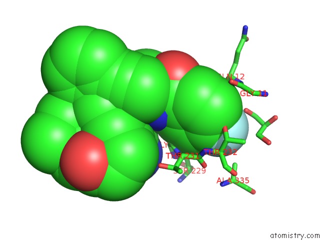
Mono view
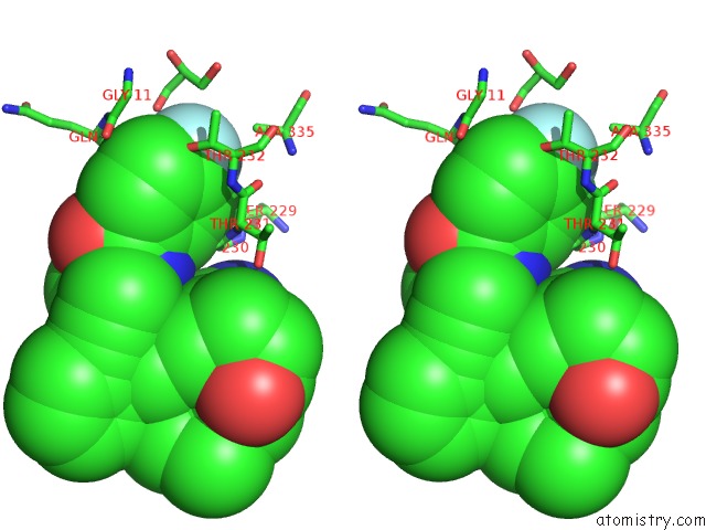
Stereo pair view
Mono view
Stereo pair view
A full contact list of Fluorine with other atoms in the F binding
site number 1 of Crystal Structure of BACE1 in Complex with Aminoquinoline Inhibitor 6 within 5.0Å range:
|
Reference:
J.B.Jordan,
D.A.Whittington,
M.D.Bartberger,
E.A.Sickmier,
K.Chen,
Y.Cheng,
T.Judd.
Fragment-Linking Approach Using (19)F uc(Nmr) Spectroscopy to Obtain Highly Potent and Selective Inhibitors of Beta-Secretase. J.Med.Chem. V. 59 3732 2016.
ISSN: ISSN 0022-2623
PubMed: 26978477
DOI: 10.1021/ACS.JMEDCHEM.5B01917
Page generated: Thu Aug 1 10:04:54 2024
ISSN: ISSN 0022-2623
PubMed: 26978477
DOI: 10.1021/ACS.JMEDCHEM.5B01917
Last articles
Zn in 9J0NZn in 9J0O
Zn in 9J0P
Zn in 9FJX
Zn in 9EKB
Zn in 9C0F
Zn in 9CAH
Zn in 9CH0
Zn in 9CH3
Zn in 9CH1