Fluorine »
PDB 5hu1-5iev »
5i56 »
Fluorine in PDB 5i56: Agonist-Bound GLUN1/GLUN2A Agonist Binding Domains with TCN201
Protein crystallography data
The structure of Agonist-Bound GLUN1/GLUN2A Agonist Binding Domains with TCN201, PDB code: 5i56
was solved by
T.-C.Mou,
S.R.Sprang,
K.B.Hansen,
with X-Ray Crystallography technique. A brief refinement statistics is given in the table below:
| Resolution Low / High (Å) | 20.03 / 2.28 |
| Space group | P 21 21 21 |
| Cell size a, b, c (Å), α, β, γ (°) | 54.259, 88.402, 126.146, 90.00, 90.00, 90.00 |
| R / Rfree (%) | 18.4 / 23.9 |
Other elements in 5i56:
The structure of Agonist-Bound GLUN1/GLUN2A Agonist Binding Domains with TCN201 also contains other interesting chemical elements:
| Chlorine | (Cl) | 1 atom |
Fluorine Binding Sites:
The binding sites of Fluorine atom in the Agonist-Bound GLUN1/GLUN2A Agonist Binding Domains with TCN201
(pdb code 5i56). This binding sites where shown within
5.0 Angstroms radius around Fluorine atom.
In total only one binding site of Fluorine was determined in the Agonist-Bound GLUN1/GLUN2A Agonist Binding Domains with TCN201, PDB code: 5i56:
In total only one binding site of Fluorine was determined in the Agonist-Bound GLUN1/GLUN2A Agonist Binding Domains with TCN201, PDB code: 5i56:
Fluorine binding site 1 out of 1 in 5i56
Go back to
Fluorine binding site 1 out
of 1 in the Agonist-Bound GLUN1/GLUN2A Agonist Binding Domains with TCN201
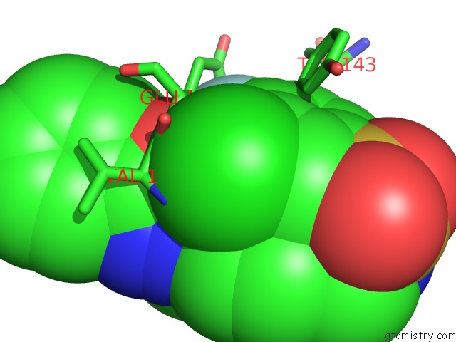
Mono view
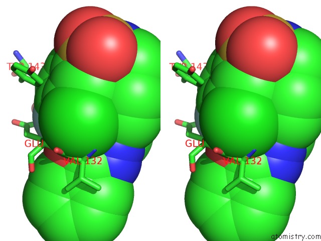
Stereo pair view
Mono view
Stereo pair view
A full contact list of Fluorine with other atoms in the F binding
site number 1 of Agonist-Bound GLUN1/GLUN2A Agonist Binding Domains with TCN201 within 5.0Å range:
|
Reference:
F.Yi,
T.C.Mou,
K.N.Dorsett,
R.A.Volkmann,
F.S.Menniti,
S.R.Sprang,
K.B.Hansen.
Structural Basis For Negative Allosteric Modulation of GLUN2A-Containing Nmda Receptors. Neuron V. 91 1316 2016.
ISSN: ISSN 0896-6273
PubMed: 27618671
DOI: 10.1016/J.NEURON.2016.08.014
Page generated: Thu Aug 1 10:06:10 2024
ISSN: ISSN 0896-6273
PubMed: 27618671
DOI: 10.1016/J.NEURON.2016.08.014
Last articles
Zn in 9J0NZn in 9J0O
Zn in 9J0P
Zn in 9FJX
Zn in 9EKB
Zn in 9C0F
Zn in 9CAH
Zn in 9CH0
Zn in 9CH3
Zn in 9CH1