Fluorine »
PDB 5ifd-5j82 »
5ifd »
Fluorine in PDB 5ifd: Crystal Structure of Polymerase Acid Protein (Pa) From Influenza A Virus, Wilson-Smith/1933 (H1N1) Bound to Follow on Fragment Ebsi-4721 1-(4-Fluorophenyl)-1H-Imidazole
Protein crystallography data
The structure of Crystal Structure of Polymerase Acid Protein (Pa) From Influenza A Virus, Wilson-Smith/1933 (H1N1) Bound to Follow on Fragment Ebsi-4721 1-(4-Fluorophenyl)-1H-Imidazole, PDB code: 5ifd
was solved by
Seattle Structural Genomics Center For Infectious Disease (Ssgcid),
with X-Ray Crystallography technique. A brief refinement statistics is given in the table below:
| Resolution Low / High (Å) | 47.78 / 2.65 |
| Space group | P 65 2 2 |
| Cell size a, b, c (Å), α, β, γ (°) | 69.030, 69.030, 397.390, 90.00, 90.00, 120.00 |
| R / Rfree (%) | 17.5 / 22.6 |
Fluorine Binding Sites:
The binding sites of Fluorine atom in the Crystal Structure of Polymerase Acid Protein (Pa) From Influenza A Virus, Wilson-Smith/1933 (H1N1) Bound to Follow on Fragment Ebsi-4721 1-(4-Fluorophenyl)-1H-Imidazole
(pdb code 5ifd). This binding sites where shown within
5.0 Angstroms radius around Fluorine atom.
In total only one binding site of Fluorine was determined in the Crystal Structure of Polymerase Acid Protein (Pa) From Influenza A Virus, Wilson-Smith/1933 (H1N1) Bound to Follow on Fragment Ebsi-4721 1-(4-Fluorophenyl)-1H-Imidazole, PDB code: 5ifd:
In total only one binding site of Fluorine was determined in the Crystal Structure of Polymerase Acid Protein (Pa) From Influenza A Virus, Wilson-Smith/1933 (H1N1) Bound to Follow on Fragment Ebsi-4721 1-(4-Fluorophenyl)-1H-Imidazole, PDB code: 5ifd:
Fluorine binding site 1 out of 1 in 5ifd
Go back to
Fluorine binding site 1 out
of 1 in the Crystal Structure of Polymerase Acid Protein (Pa) From Influenza A Virus, Wilson-Smith/1933 (H1N1) Bound to Follow on Fragment Ebsi-4721 1-(4-Fluorophenyl)-1H-Imidazole

Mono view
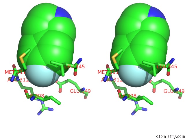
Stereo pair view
Mono view
Stereo pair view
A full contact list of Fluorine with other atoms in the F binding
site number 1 of Crystal Structure of Polymerase Acid Protein (Pa) From Influenza A Virus, Wilson-Smith/1933 (H1N1) Bound to Follow on Fragment Ebsi-4721 1-(4-Fluorophenyl)-1H-Imidazole within 5.0Å range:
|
Reference:
P.Pierce,
M.M.Muruthi,
J.Abendroth,
S.O.Moen,
D.W.Begley,
D.R.Davies,
V.M.Marathias,
B.L.Staker,
P.J.Myler,
D.D.Lorimer,
T.E.Edwards.
Fragment Screening By Std uc(Nmr) Identifies Novel Site Binders Against Influenza A Virus Polymerase Pa To Be Published.
Page generated: Thu Aug 1 10:16:19 2024
Last articles
Zn in 9MJ5Zn in 9HNW
Zn in 9G0L
Zn in 9FNE
Zn in 9DZN
Zn in 9E0I
Zn in 9D32
Zn in 9DAK
Zn in 8ZXC
Zn in 8ZUF