Fluorine »
PDB 5ifd-5j82 »
5ikv »
Fluorine in PDB 5ikv: The Structure of Flufenamic Acid Bound to Human Cyclooxygenase-2
Enzymatic activity of The Structure of Flufenamic Acid Bound to Human Cyclooxygenase-2
All present enzymatic activity of The Structure of Flufenamic Acid Bound to Human Cyclooxygenase-2:
1.14.99.1;
1.14.99.1;
Protein crystallography data
The structure of The Structure of Flufenamic Acid Bound to Human Cyclooxygenase-2, PDB code: 5ikv
was solved by
B.J.Orlando,
M.G.Malkowski,
with X-Ray Crystallography technique. A brief refinement statistics is given in the table below:
| Resolution Low / High (Å) | 30.01 / 2.51 |
| Space group | I 2 2 2 |
| Cell size a, b, c (Å), α, β, γ (°) | 126.918, 149.334, 184.766, 90.00, 90.00, 90.00 |
| R / Rfree (%) | 18.5 / 22.4 |
Other elements in 5ikv:
The structure of The Structure of Flufenamic Acid Bound to Human Cyclooxygenase-2 also contains other interesting chemical elements:
| Cobalt | (Co) | 2 atoms |
Fluorine Binding Sites:
The binding sites of Fluorine atom in the The Structure of Flufenamic Acid Bound to Human Cyclooxygenase-2
(pdb code 5ikv). This binding sites where shown within
5.0 Angstroms radius around Fluorine atom.
In total 6 binding sites of Fluorine where determined in the The Structure of Flufenamic Acid Bound to Human Cyclooxygenase-2, PDB code: 5ikv:
Jump to Fluorine binding site number: 1; 2; 3; 4; 5; 6;
In total 6 binding sites of Fluorine where determined in the The Structure of Flufenamic Acid Bound to Human Cyclooxygenase-2, PDB code: 5ikv:
Jump to Fluorine binding site number: 1; 2; 3; 4; 5; 6;
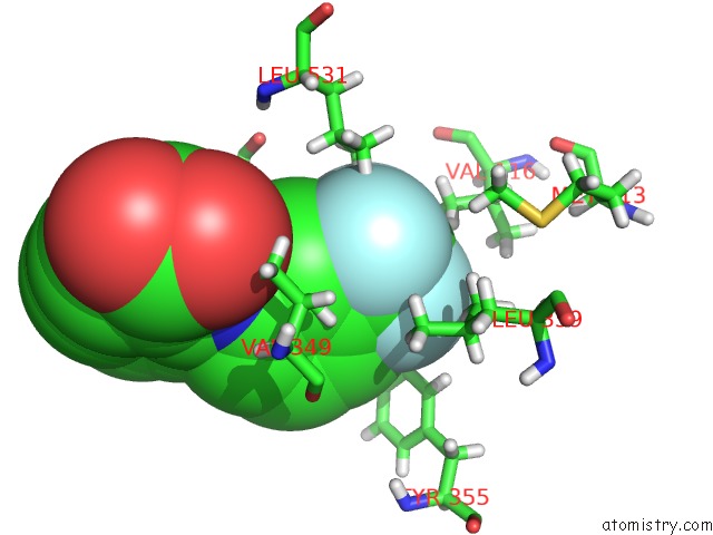
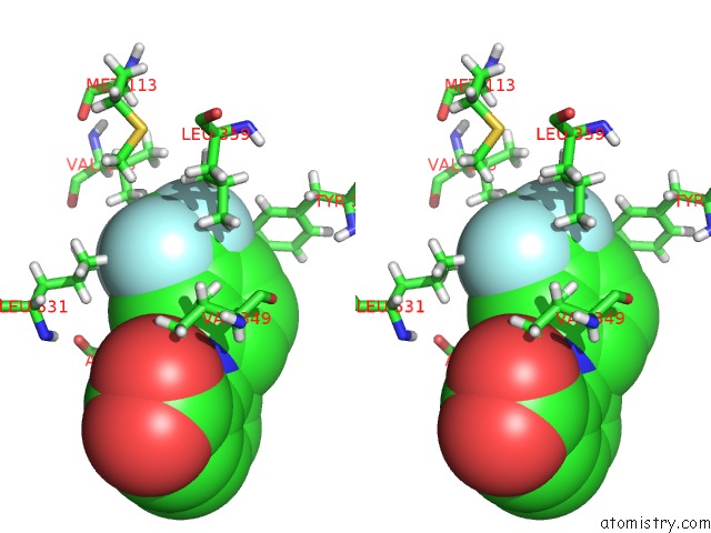
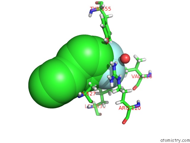
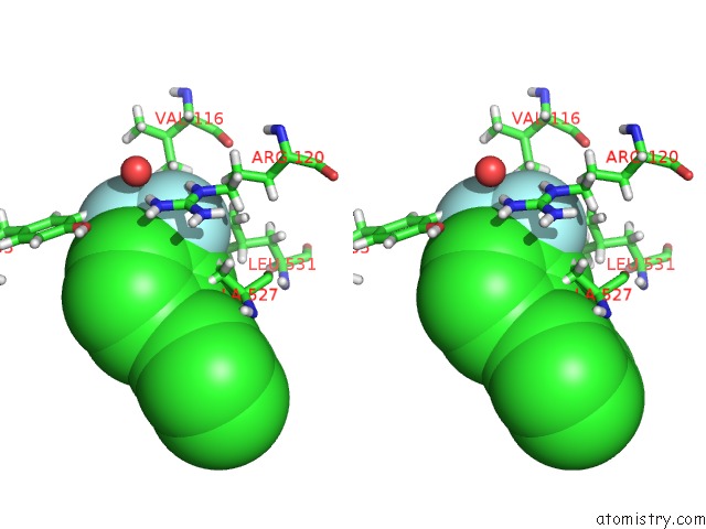
Fluorine binding site 1 out of 6 in 5ikv
Go back to
Fluorine binding site 1 out
of 6 in the The Structure of Flufenamic Acid Bound to Human Cyclooxygenase-2
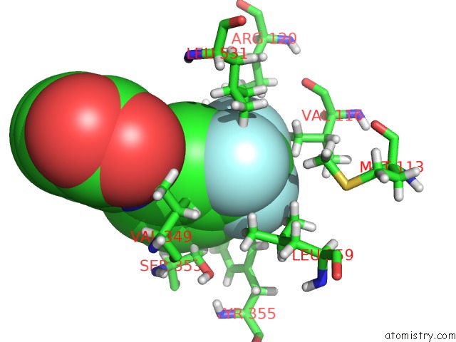
Mono view
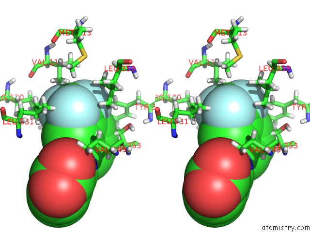
Stereo pair view
Mono view
Stereo pair view
A full contact list of Fluorine with other atoms in the F binding
site number 1 of The Structure of Flufenamic Acid Bound to Human Cyclooxygenase-2 within 5.0Å range:
|
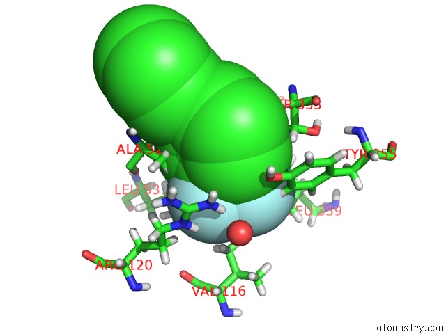
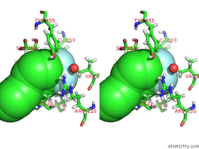
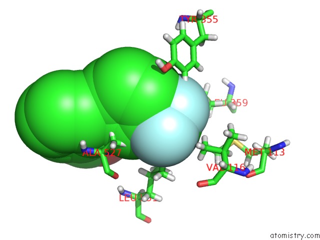
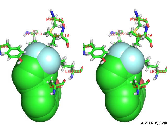
Fluorine binding site 2 out of 6 in 5ikv
Go back to
Fluorine binding site 2 out
of 6 in the The Structure of Flufenamic Acid Bound to Human Cyclooxygenase-2
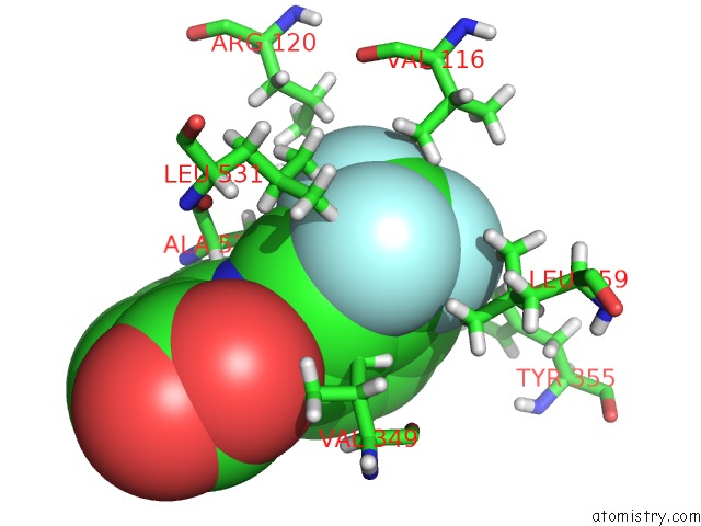
Mono view
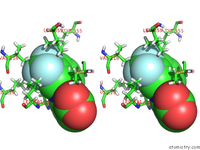
Stereo pair view
Mono view
Stereo pair view
A full contact list of Fluorine with other atoms in the F binding
site number 2 of The Structure of Flufenamic Acid Bound to Human Cyclooxygenase-2 within 5.0Å range:
|
Fluorine binding site 3 out of 6 in 5ikv
Go back to
Fluorine binding site 3 out
of 6 in the The Structure of Flufenamic Acid Bound to Human Cyclooxygenase-2
Mono view
Stereo pair view
Mono view
Stereo pair view
A full contact list of Fluorine with other atoms in the F binding
site number 3 of The Structure of Flufenamic Acid Bound to Human Cyclooxygenase-2 within 5.0Å range:
|
Fluorine binding site 4 out of 6 in 5ikv
Go back to
Fluorine binding site 4 out
of 6 in the The Structure of Flufenamic Acid Bound to Human Cyclooxygenase-2
Mono view
Stereo pair view
Mono view
Stereo pair view
A full contact list of Fluorine with other atoms in the F binding
site number 4 of The Structure of Flufenamic Acid Bound to Human Cyclooxygenase-2 within 5.0Å range:
|
Fluorine binding site 5 out of 6 in 5ikv
Go back to
Fluorine binding site 5 out
of 6 in the The Structure of Flufenamic Acid Bound to Human Cyclooxygenase-2
Mono view
Stereo pair view
Mono view
Stereo pair view
A full contact list of Fluorine with other atoms in the F binding
site number 5 of The Structure of Flufenamic Acid Bound to Human Cyclooxygenase-2 within 5.0Å range:
|
Fluorine binding site 6 out of 6 in 5ikv
Go back to
Fluorine binding site 6 out
of 6 in the The Structure of Flufenamic Acid Bound to Human Cyclooxygenase-2
Mono view
Stereo pair view
Mono view
Stereo pair view
A full contact list of Fluorine with other atoms in the F binding
site number 6 of The Structure of Flufenamic Acid Bound to Human Cyclooxygenase-2 within 5.0Å range:
|
Reference:
B.J.Orlando,
M.G.Malkowski.
Substrate-Selective Inhibition of Cyclooxygeanse-2 By Fenamic Acid Derivatives Is Dependent on Peroxide Tone. J.Biol.Chem. V. 291 15069 2016.
ISSN: ESSN 1083-351X
PubMed: 27226593
DOI: 10.1074/JBC.M116.725713
Page generated: Thu Aug 1 10:16:19 2024
ISSN: ESSN 1083-351X
PubMed: 27226593
DOI: 10.1074/JBC.M116.725713
Last articles
Ca in 5OC9Ca in 5ODC
Ca in 5O8N
Ca in 5O8X
Ca in 5O32
Ca in 5O76
Ca in 5O8L
Ca in 5O7W
Ca in 5O7E
Ca in 5O7U