Fluorine »
PDB 5ifd-5j82 »
5j82 »
Fluorine in PDB 5j82: Crystal Structure of HSP90-Alpha N-Domain in Complex 5-[4-(2-Fluoro- Phenyl)-5-Oxo-4,5-Dihydro-1H-[1,2,4]Triazol-3-Yl]-2,4-Dihydroxy-N- Isopropyl-N-Methyl-Benzenesulfonamide
Protein crystallography data
The structure of Crystal Structure of HSP90-Alpha N-Domain in Complex 5-[4-(2-Fluoro- Phenyl)-5-Oxo-4,5-Dihydro-1H-[1,2,4]Triazol-3-Yl]-2,4-Dihydroxy-N- Isopropyl-N-Methyl-Benzenesulfonamide, PDB code: 5j82
was solved by
M.Amaral,
P.Matias,
with X-Ray Crystallography technique. A brief refinement statistics is given in the table below:
| Resolution Low / High (Å) | 36.59 / 2.17 |
| Space group | I 2 2 2 |
| Cell size a, b, c (Å), α, β, γ (°) | 67.696, 90.922, 99.028, 90.00, 90.00, 90.00 |
| R / Rfree (%) | 23.4 / 27.6 |
Fluorine Binding Sites:
The binding sites of Fluorine atom in the Crystal Structure of HSP90-Alpha N-Domain in Complex 5-[4-(2-Fluoro- Phenyl)-5-Oxo-4,5-Dihydro-1H-[1,2,4]Triazol-3-Yl]-2,4-Dihydroxy-N- Isopropyl-N-Methyl-Benzenesulfonamide
(pdb code 5j82). This binding sites where shown within
5.0 Angstroms radius around Fluorine atom.
In total only one binding site of Fluorine was determined in the Crystal Structure of HSP90-Alpha N-Domain in Complex 5-[4-(2-Fluoro- Phenyl)-5-Oxo-4,5-Dihydro-1H-[1,2,4]Triazol-3-Yl]-2,4-Dihydroxy-N- Isopropyl-N-Methyl-Benzenesulfonamide, PDB code: 5j82:
In total only one binding site of Fluorine was determined in the Crystal Structure of HSP90-Alpha N-Domain in Complex 5-[4-(2-Fluoro- Phenyl)-5-Oxo-4,5-Dihydro-1H-[1,2,4]Triazol-3-Yl]-2,4-Dihydroxy-N- Isopropyl-N-Methyl-Benzenesulfonamide, PDB code: 5j82:
Fluorine binding site 1 out of 1 in 5j82
Go back to
Fluorine binding site 1 out
of 1 in the Crystal Structure of HSP90-Alpha N-Domain in Complex 5-[4-(2-Fluoro- Phenyl)-5-Oxo-4,5-Dihydro-1H-[1,2,4]Triazol-3-Yl]-2,4-Dihydroxy-N- Isopropyl-N-Methyl-Benzenesulfonamide
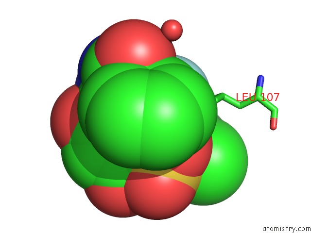
Mono view
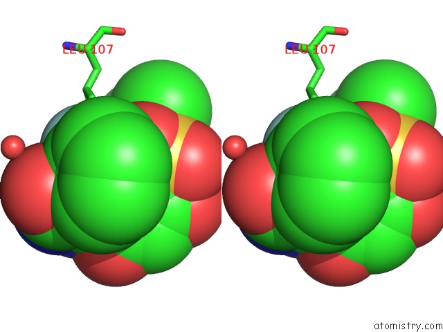
Stereo pair view
Mono view
Stereo pair view
A full contact list of Fluorine with other atoms in the F binding
site number 1 of Crystal Structure of HSP90-Alpha N-Domain in Complex 5-[4-(2-Fluoro- Phenyl)-5-Oxo-4,5-Dihydro-1H-[1,2,4]Triazol-3-Yl]-2,4-Dihydroxy-N- Isopropyl-N-Methyl-Benzenesulfonamide within 5.0Å range:
|
Reference:
M.Amaral,
D.B.Kokh,
J.Bomke,
A.Wegener,
H.P.Buchstaller,
H.M.Eggenweiler,
P.Matias,
C.Sirrenberg,
R.C.Wade,
M.Frech.
Protein Conformational Flexibility Modulates Kinetics and Thermodynamics of Drug Binding. Nat Commun V. 8 2276 2017.
ISSN: ESSN 2041-1723
PubMed: 29273709
DOI: 10.1038/S41467-017-02258-W
Page generated: Thu Aug 1 10:28:00 2024
ISSN: ESSN 2041-1723
PubMed: 29273709
DOI: 10.1038/S41467-017-02258-W
Last articles
Zn in 9MJ5Zn in 9HNW
Zn in 9G0L
Zn in 9FNE
Zn in 9DZN
Zn in 9E0I
Zn in 9D32
Zn in 9DAK
Zn in 8ZXC
Zn in 8ZUF