Atomistry » Fluorine » PDB 5lck-5lz4 » 5lih
Atomistry »
  Fluorine »
    PDB 5lck-5lz4 »
      5lih »

Fluorine in PDB 5lih: Structure of A Peptide-Substrate Bound to Pkciota Core Kinase Domain

Enzymatic activity of Structure of A Peptide-Substrate Bound to Pkciota Core Kinase Domain

All present enzymatic activity of Structure of A Peptide-Substrate Bound to Pkciota Core Kinase Domain:
2.7.11.13;

Protein crystallography data

The structure of Structure of A Peptide-Substrate Bound to Pkciota Core Kinase Domain, PDB code: 5lih was solved by E.V.Soriano, A.G.Purkiss, N.Q.Mcdonald, with X-Ray Crystallography technique. A brief refinement statistics is given in the table below:

Resolution Low / High (Å) 67.28 / 3.25
Space group P 21 21 21
Cell size a, b, c (Å), α, β, γ (°) 78.980, 84.230, 111.830, 90.00, 90.00, 90.00
R / Rfree (%) 25.7 / 28.4

Other elements in 5lih:

The structure of Structure of A Peptide-Substrate Bound to Pkciota Core Kinase Domain also contains other interesting chemical elements:

Aluminium (Al) 4 atoms
Manganese (Mn) 5 atoms

Fluorine Binding Sites:

Pages:

>>> Page 1 <<< Page 2, Binding sites: 11 - 12;

Binding sites:

The binding sites of Fluorine atom in the Structure of A Peptide-Substrate Bound to Pkciota Core Kinase Domain (pdb code 5lih). This binding sites where shown within 5.0 Angstroms radius around Fluorine atom.
In total 12 binding sites of Fluorine where determined in the Structure of A Peptide-Substrate Bound to Pkciota Core Kinase Domain, PDB code: 5lih:
Jump to Fluorine binding site number: 1; 2; 3; 4; 5; 6; 7; 8; 9; 10;

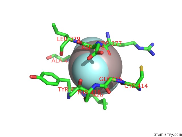
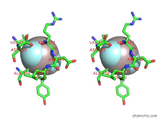
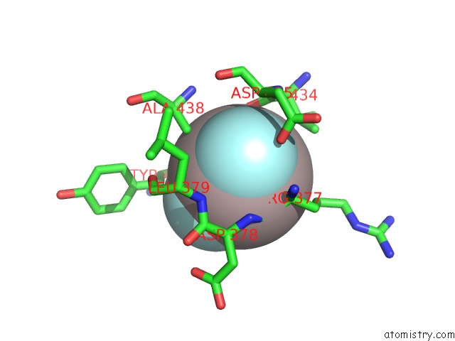
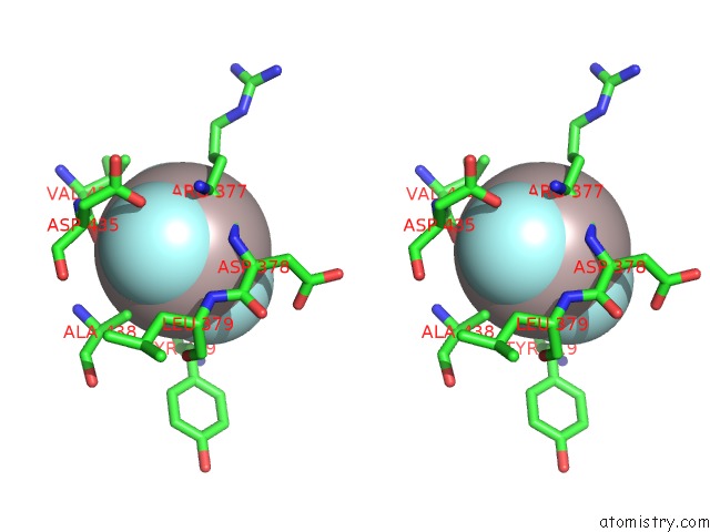
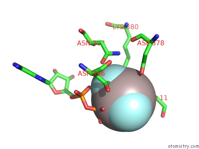
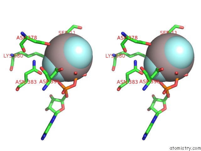
Fluorine binding site 1 out of 12 in 5lih

Go back to Fluorine Binding Sites List in 5lih
Fluorine binding site 1 out of 12 in the Structure of A Peptide-Substrate Bound to Pkciota Core Kinase Domain


Mono view


Stereo pair view

A full contact list of Fluorine with other atoms in the F binding site number 1 of Structure of A Peptide-Substrate Bound to Pkciota Core Kinase Domain within 5.0Å range:
probe atom residue distance (Å) B Occ
A:F602

b:37.9
occ:1.00
F1 A:AF3602 0.0 37.9 1.0
AL A:AF3602 1.8 37.9 1.0
F3 A:AF3602 3.1 37.9 1.0
F2 A:AF3602 3.1 37.9 1.0
O A:ARG377 3.5 31.5 1.0
N A:THR416 3.5 32.8 1.0
CA A:GLY415 3.6 34.2 1.0
O A:TYR419 3.7 29.4 1.0
CB A:TYR419 3.7 29.8 1.0
C A:ARG377 3.8 31.8 1.0
CA A:ASP378 3.9 30.1 1.0
C A:TYR419 3.9 29.4 1.0
N A:ASP378 4.0 30.6 1.0
C A:GLY415 4.1 34.1 1.0
N A:ILE420 4.3 30.8 1.0
O A:THR416 4.3 33.0 1.0
CA A:TYR419 4.5 29.9 1.0
CA A:ILE420 4.5 31.1 1.0
N A:LEU379 4.5 28.6 1.0
C A:ASP378 4.5 30.3 1.0
CA A:THR416 4.6 33.4 1.0
CA A:ARG377 4.6 33.3 1.0
O A:CYS414 4.7 35.9 1.0
CB A:ALA438 4.7 27.8 1.0
CG1 A:ILE420 4.7 31.3 1.0
OD1 A:ASP378 4.8 30.5 1.0
N A:GLY415 4.8 34.7 1.0
CB A:THR416 4.8 34.2 1.0
O A:LEU379 5.0 28.1 1.0
CG A:TYR419 5.0 31.1 1.0
C A:THR416 5.0 33.4 1.0

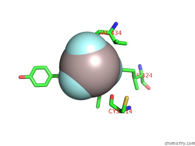
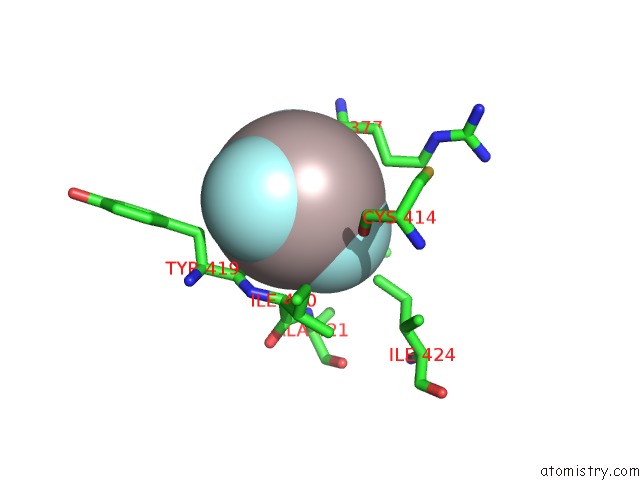
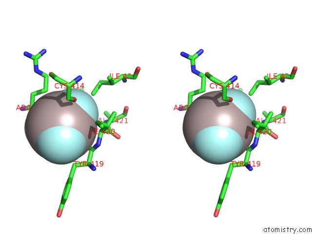
Fluorine binding site 2 out of 12 in 5lih

Go back to Fluorine Binding Sites List in 5lih
Fluorine binding site 2 out of 12 in the Structure of A Peptide-Substrate Bound to Pkciota Core Kinase Domain


Mono view


Stereo pair view

A full contact list of Fluorine with other atoms in the F binding site number 2 of Structure of A Peptide-Substrate Bound to Pkciota Core Kinase Domain within 5.0Å range:
probe atom residue distance (Å) B Occ
A:F602

b:37.9
occ:1.00
F2 A:AF3602 0.0 37.9 1.0
AL A:AF3602 1.8 37.9 1.0
O A:VAL434 2.7 31.7 1.0
F3 A:AF3602 3.1 37.9 1.0
F1 A:AF3602 3.1 37.9 1.0
CB A:ALA438 3.4 27.8 1.0
C A:VAL434 3.5 33.1 1.0
CA A:ARG377 3.7 33.3 1.0
OD1 A:ASP435 3.8 31.5 1.0
C A:ARG377 3.9 31.8 1.0
CA A:ASP435 4.0 31.6 1.0
N A:ASP435 4.1 32.0 1.0
CB A:VAL434 4.1 35.1 1.0
N A:ASP378 4.1 30.6 1.0
CG2 A:VAL434 4.2 35.9 1.0
O A:TYR419 4.3 29.4 1.0
N A:ARG377 4.3 34.3 1.0
O A:ARG377 4.4 31.5 1.0
CA A:VAL434 4.4 34.6 1.0
N A:LEU379 4.6 28.6 1.0
N A:ALA438 4.6 28.4 1.0
CA A:ALA438 4.6 27.9 1.0
CG A:ASP435 4.7 32.4 1.0
CB A:ARG377 4.8 34.8 1.0
CA A:ASP378 4.9 30.1 1.0
C A:ASP435 4.9 31.6 1.0

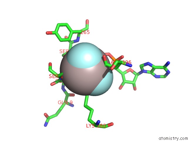
Fluorine binding site 3 out of 12 in 5lih

Go back to Fluorine Binding Sites List in 5lih
Fluorine binding site 3 out of 12 in the Structure of A Peptide-Substrate Bound to Pkciota Core Kinase Domain


Mono view


Stereo pair view

A full contact list of Fluorine with other atoms in the F binding site number 3 of Structure of A Peptide-Substrate Bound to Pkciota Core Kinase Domain within 5.0Å range:
probe atom residue distance (Å) B Occ
A:F602

b:37.9
occ:1.00
F3 A:AF3602 0.0 37.9 1.0
AL A:AF3602 1.8 37.9 1.0
F2 A:AF3602 3.1 37.9 1.0
CD1 A:ILE424 3.1 35.2 1.0
F1 A:AF3602 3.1 37.9 1.0
N A:ALA421 3.2 26.0 1.0
CA A:ILE420 3.2 31.1 1.0
O A:TYR419 3.4 29.4 1.0
C A:ILE420 3.7 31.6 1.0
N A:ILE420 4.0 30.8 1.0
C A:TYR419 4.0 29.4 1.0
CB A:ILE420 4.1 31.5 1.0
CB A:VAL434 4.1 35.1 1.0
CG1 A:ILE420 4.2 31.3 1.0
O A:CYS414 4.2 35.9 1.0
CB A:ALA421 4.3 37.2 1.0
CA A:ALA421 4.3 36.7 1.0
O A:VAL434 4.3 31.7 1.0
CG2 A:ILE420 4.4 32.3 1.0
CG1 A:ILE424 4.6 36.4 1.0
CG1 A:VAL434 4.7 37.0 1.0
CG2 A:VAL434 4.8 35.9 1.0
O A:ILE420 4.9 31.8 1.0
CA A:VAL434 4.9 34.6 1.0
C A:VAL434 5.0 33.1 1.0

Fluorine binding site 4 out of 12 in 5lih

Go back to Fluorine Binding Sites List in 5lih
Fluorine binding site 4 out of 12 in the Structure of A Peptide-Substrate Bound to Pkciota Core Kinase Domain


Mono view


Stereo pair view

A full contact list of Fluorine with other atoms in the F binding site number 4 of Structure of A Peptide-Substrate Bound to Pkciota Core Kinase Domain within 5.0Å range:
probe atom residue distance (Å) B Occ
A:F605

b:22.1
occ:1.00
F1 A:AF3605 0.0 22.1 1.0
AL A:AF3605 1.8 54.5 1.0
MN A:MN603 2.0 31.8 1.0
OD2 A:ASP396 2.5 34.3 1.0
O1B A:ADP601 2.5 40.8 1.0
O1A A:ADP601 3.0 33.5 1.0
OD1 A:ASN383 3.0 31.2 0.5
F3 A:AF3605 3.1 34.8 1.0
F2 A:AF3605 3.1 63.9 1.0
CE A:LYS380 3.2 31.4 1.0
CG A:ASP396 3.7 34.4 1.0
PB A:ADP601 3.8 45.0 1.0
CG A:ASN383 3.9 30.5 0.5
ND2 A:ASN383 3.9 29.8 0.5
O2B A:ADP601 4.0 43.8 1.0
PA A:ADP601 4.3 33.9 1.0
MN A:MN604 4.3 38.7 1.0
OD2 A:ASP378 4.4 30.9 1.0
CB A:ASP396 4.5 34.8 1.0
O3A A:ADP601 4.5 36.4 1.0
OD1 A:ASP396 4.5 34.5 1.0
CD A:LYS380 4.7 31.2 1.0
OG F:SER11 4.7 43.9 1.0
CB A:ASP382 4.8 36.9 1.0
O3B A:ADP601 5.0 58.6 1.0

Fluorine binding site 5 out of 12 in 5lih

Go back to Fluorine Binding Sites List in 5lih
Fluorine binding site 5 out of 12 in the Structure of A Peptide-Substrate Bound to Pkciota Core Kinase Domain


Mono view


Stereo pair view

A full contact list of Fluorine with other atoms in the F binding site number 5 of Structure of A Peptide-Substrate Bound to Pkciota Core Kinase Domain within 5.0Å range:
probe atom residue distance (Å) B Occ
A:F605

b:63.9
occ:1.00
F2 A:AF3605 0.0 63.9 1.0
AL A:AF3605 1.8 54.5 1.0
O1B A:ADP601 2.8 40.8 1.0
OG A:SER264 2.9 37.6 1.0
N A:SER264 3.0 36.9 1.0
F1 A:AF3605 3.1 22.1 1.0
F3 A:AF3605 3.1 34.8 1.0
N F:SER11 3.2 41.5 1.0
CA F:GLY10 3.5 40.5 1.0
OG F:SER11 3.6 43.9 1.0
CB A:SER264 3.7 38.0 1.0
PB A:ADP601 3.8 45.0 1.0
CA A:GLY263 3.8 36.4 1.0
C A:GLY263 3.9 37.1 1.0
C F:GLY10 3.9 40.8 1.0
CA A:SER264 3.9 37.8 1.0
O3B A:ADP601 4.1 58.6 1.0
CB F:SER11 4.2 43.2 1.0
O2B A:ADP601 4.3 43.8 1.0
CA F:SER11 4.3 42.2 1.0
O F:GLN9 4.5 39.6 1.0
MN A:MN603 4.6 31.8 1.0
N F:GLY10 4.7 39.7 1.0
N A:TYR265 4.7 35.5 1.0
OD2 A:ASP396 4.7 34.3 1.0
C A:SER264 4.8 38.3 1.0
CE A:LYS380 4.9 31.4 1.0
O F:SER11 4.9 43.0 1.0
MN A:MN604 5.0 38.7 1.0

Fluorine binding site 6 out of 12 in 5lih

Go back to Fluorine Binding Sites List in 5lih
Fluorine binding site 6 out of 12 in the Structure of A Peptide-Substrate Bound to Pkciota Core Kinase Domain


Mono view


Stereo pair view

A full contact list of Fluorine with other atoms in the F binding site number 6 of Structure of A Peptide-Substrate Bound to Pkciota Core Kinase Domain within 5.0Å range:
probe atom residue distance (Å) B Occ
A:F605

b:34.8
occ:1.00
F3 A:AF3605 0.0 34.8 1.0
AL A:AF3605 1.8 54.5 1.0
MN A:MN604 2.0 38.7 1.0
O2B A:ADP601 2.6 43.8 1.0
OG F:SER11 2.6 43.9 1.0
OD2 A:ASP396 2.8 34.3 1.0
F1 A:AF3605 3.1 22.1 1.0
F2 A:AF3605 3.1 63.9 1.0
O1B A:ADP601 3.2 40.8 1.0
PB A:ADP601 3.4 45.0 1.0
OD1 A:ASP396 3.5 34.5 1.0
CG A:ASP396 3.5 34.4 1.0
CB F:SER11 3.6 43.2 1.0
OD2 A:ASP378 3.7 30.9 1.0
OG A:SER264 4.1 37.6 1.0
CD2 A:TYR265 4.2 36.2 1.0
O3B A:ADP601 4.2 58.6 1.0
N F:SER11 4.6 41.5 1.0
O1A A:ADP601 4.6 33.5 1.0
MN A:MN603 4.6 31.8 1.0
O3A A:ADP601 4.7 36.4 1.0
CA F:SER11 4.7 42.2 1.0
CG A:ASP378 4.9 30.4 1.0
CE2 A:TYR265 4.9 36.9 1.0
CE A:MET399 4.9 35.9 1.0
CB A:ASP396 5.0 34.8 1.0

Fluorine binding site 7 out of 12 in 5lih

Go back to Fluorine Binding Sites List in 5lih
Fluorine binding site 7 out of 12 in the Structure of A Peptide-Substrate Bound to Pkciota Core Kinase Domain


Mono view


Stereo pair view

A full contact list of Fluorine with other atoms in the F binding site number 7 of Structure of A Peptide-Substrate Bound to Pkciota Core Kinase Domain within 5.0Å range:
probe atom residue distance (Å) B Occ
B:F602

b:36.8
occ:1.00
F1 B:AF3602 0.0 36.8 1.0
AL B:AF3602 1.8 36.8 1.0
F3 B:AF3602 3.1 36.8 1.0
F2 B:AF3602 3.1 36.8 1.0
CB B:TYR419 3.4 39.0 1.0
O B:TYR419 3.5 40.7 1.0
C B:TYR419 3.7 41.0 1.0
O B:ARG377 3.8 43.1 1.0
N B:THR416 3.8 43.0 1.0
CA B:GLY415 3.9 44.9 1.0
CA B:ASP378 4.0 40.5 1.0
C B:ARG377 4.1 42.0 1.0
N B:ILE420 4.1 36.6 1.0
CA B:TYR419 4.1 39.7 1.0
N B:ASP378 4.2 40.7 1.0
O B:THR416 4.4 42.8 1.0
C B:GLY415 4.4 45.6 1.0
CA B:ILE420 4.4 37.5 1.0
N B:LEU379 4.4 35.6 1.0
CB B:ALA438 4.5 31.6 1.0
C B:ASP378 4.5 39.5 1.0
CG B:TYR419 4.6 36.0 1.0
O B:LEU379 4.7 34.5 1.0
OD1 B:ASP378 4.8 41.5 1.0
CA B:THR416 4.8 42.9 1.0
CG1 B:ILE420 4.9 38.8 1.0
CA B:ARG377 4.9 42.5 1.0
CB B:THR416 4.9 40.0 1.0
N B:TYR419 5.0 38.6 1.0
O B:CYS414 5.0 43.8 1.0

Fluorine binding site 8 out of 12 in 5lih

Go back to Fluorine Binding Sites List in 5lih
Fluorine binding site 8 out of 12 in the Structure of A Peptide-Substrate Bound to Pkciota Core Kinase Domain


Mono view


Stereo pair view

A full contact list of Fluorine with other atoms in the F binding site number 8 of Structure of A Peptide-Substrate Bound to Pkciota Core Kinase Domain within 5.0Å range:
probe atom residue distance (Å) B Occ
B:F602

b:36.8
occ:1.00
F2 B:AF3602 0.0 36.8 1.0
AL B:AF3602 1.8 36.8 1.0
F3 B:AF3602 3.1 36.8 1.0
F1 B:AF3602 3.1 36.8 1.0
O B:VAL434 3.2 35.1 1.0
CA B:ARG377 3.3 42.5 1.0
C B:ARG377 3.3 42.0 1.0
OD1 B:ASP435 3.4 33.8 1.0
N B:ASP378 3.5 40.7 1.0
CB B:ALA438 3.5 31.6 1.0
C B:VAL434 3.8 35.1 1.0
N B:ARG377 3.8 41.3 1.0
N B:LEU379 3.9 35.6 1.0
O B:ARG377 4.0 43.1 1.0
CA B:ASP435 4.0 33.0 1.0
N B:ASP435 4.2 33.7 1.0
CA B:ASP378 4.2 40.5 1.0
CG2 B:VAL434 4.4 36.9 1.0
CG B:ASP435 4.4 34.4 1.0
CB B:ARG377 4.5 44.2 1.0
CB B:VAL434 4.5 37.3 1.0
C B:ASP378 4.6 39.5 1.0
CG B:ARG377 4.6 46.0 1.0
CB B:LEU379 4.7 35.3 1.0
CA B:ALA438 4.8 31.5 1.0
CA B:VAL434 4.8 36.5 1.0
O B:TYR419 4.8 40.7 1.0
CB B:ASP435 4.8 33.9 1.0
CA B:LEU379 4.9 35.3 1.0
N B:ALA438 4.9 31.9 1.0
C B:ASP435 5.0 32.5 1.0

Fluorine binding site 9 out of 12 in 5lih

Go back to Fluorine Binding Sites List in 5lih
Fluorine binding site 9 out of 12 in the Structure of A Peptide-Substrate Bound to Pkciota Core Kinase Domain


Mono view


Stereo pair view

A full contact list of Fluorine with other atoms in the F binding site number 9 of Structure of A Peptide-Substrate Bound to Pkciota Core Kinase Domain within 5.0Å range:
probe atom residue distance (Å) B Occ
B:F602

b:36.8
occ:1.00
F3 B:AF3602 0.0 36.8 1.0
AL B:AF3602 1.8 36.8 1.0
F2 B:AF3602 3.1 36.8 1.0
F1 B:AF3602 3.1 36.8 1.0
CD1 B:ILE424 3.3 42.0 1.0
CB B:VAL434 3.6 37.3 1.0
O B:TYR419 3.9 40.7 1.0
N B:ALA421 4.0 35.5 1.0
CA B:ILE420 4.0 37.5 1.0
O B:VAL434 4.0 35.1 1.0
CG B:ARG377 4.0 46.0 1.0
CG2 B:VAL434 4.1 36.9 1.0
O B:CYS414 4.2 43.8 1.0
CG1 B:VAL434 4.4 38.0 1.0
O B:ARG377 4.4 43.1 1.0
C B:ILE420 4.5 37.8 1.0
C B:VAL434 4.5 35.1 1.0
CA B:VAL434 4.6 36.5 1.0
CA B:ARG377 4.6 42.5 1.0
C B:TYR419 4.6 41.0 1.0
C B:ARG377 4.6 42.0 1.0
N B:ILE420 4.7 36.6 1.0
CG1 B:ILE420 4.8 38.8 1.0
CB B:ALA421 4.8 36.3 1.0
CG1 B:ILE424 4.8 42.9 1.0
CB B:ILE420 4.9 38.7 1.0
CB B:ARG377 4.9 44.2 1.0
CA B:ALA421 5.0 36.0 1.0

Fluorine binding site 10 out of 12 in 5lih

Go back to Fluorine Binding Sites List in 5lih
Fluorine binding site 10 out of 12 in the Structure of A Peptide-Substrate Bound to Pkciota Core Kinase Domain


Mono view


Stereo pair view

A full contact list of Fluorine with other atoms in the F binding site number 10 of Structure of A Peptide-Substrate Bound to Pkciota Core Kinase Domain within 5.0Å range:
probe atom residue distance (Å) B Occ
B:F605

b:26.9
occ:1.00
F1 B:AF3605 0.0 26.9 1.0
AL B:AF3605 1.8 59.4 1.0
MN B:MN603 1.9 50.6 1.0
O1B B:ADP601 2.1 44.1 1.0
OD2 B:ASP396 2.4 41.9 1.0
MN B:MN604 2.9 40.7 1.0
F3 B:AF3605 3.1 39.6 1.0
F2 B:AF3605 3.1 68.7 1.0
O1A B:ADP601 3.1 46.0 1.0
CE B:LYS380 3.4 39.0 1.0
PB B:ADP601 3.5 38.5 1.0
OD1 B:ASN383 3.6 37.9 1.0
CG B:ASP396 3.6 41.8 1.0
O3B B:ADP601 4.0 46.1 1.0
OD2 B:ASP378 4.3 41.2 1.0
PA B:ADP601 4.3 49.1 1.0
O3A B:ADP601 4.3 45.9 1.0
OD1 B:ASP396 4.4 42.6 1.0
CG B:ASN383 4.4 37.1 1.0
ND2 B:ASN383 4.4 36.5 1.0
O2B B:ADP601 4.5 38.3 1.0
OG G:SER11 4.5 48.4 1.0
CB B:ASP396 4.6 41.6 1.0
CB G:SER11 4.7 46.9 1.0
N G:SER11 5.0 44.5 1.0
CD B:LYS380 5.0 38.5 1.0

Reference:

E.V.Soriano, M.E.Ivanova, G.Fletcher, P.Riou, P.P.Knowles, K.Barnouin, A.Purkiss, B.Kostelecky, P.Saiu, M.Linch, A.Elbediwy, S.Kjr, N.O'reilly, A.P.Snijders, P.J.Parker, B.J.Thompson, N.Q.Mcdonald. Apkc Inhibition By PAR3 CR3 Flanking Regions Controls Substrate Access and Underpins Apical-Junctional Polarization. Dev.Cell V. 38 384 2016.
ISSN: ISSN 1534-5807
PubMed: 27554858
DOI: 10.1016/J.DEVCEL.2016.07.018
Page generated: Sun Dec 13 12:27:06 2020

Last articles

Cl in 6COG
Cl in 6CST
Cl in 6CTW
Cl in 6CTZ
Cl in 6CTT
Cl in 6CSP
Cl in 6CU9
Cl in 6CTX
Cl in 6CTU
Cl in 6CTP
© Copyright 2008-2020 by atomistry.com
Home   |    Site Map   |    Copyright   |    Contact us   |    Privacy