Fluorine »
PDB 5msb-5njh »
5n2f »
Fluorine in PDB 5n2f: Structure of Pd-L1/Small-Molecule Inhibitor Complex
Protein crystallography data
The structure of Structure of Pd-L1/Small-Molecule Inhibitor Complex, PDB code: 5n2f
was solved by
K.Guzik,
K.M.Zak,
P.Grudnik,
G.Dubin,
T.A.Holak,
with X-Ray Crystallography technique. A brief refinement statistics is given in the table below:
| Resolution Low / High (Å) | 36.84 / 1.70 |
| Space group | P 2 21 21 |
| Cell size a, b, c (Å), α, β, γ (°) | 51.815, 52.379, 111.526, 90.00, 90.00, 90.00 |
| R / Rfree (%) | 19.1 / 23 |
Fluorine Binding Sites:
The binding sites of Fluorine atom in the Structure of Pd-L1/Small-Molecule Inhibitor Complex
(pdb code 5n2f). This binding sites where shown within
5.0 Angstroms radius around Fluorine atom.
In total 2 binding sites of Fluorine where determined in the Structure of Pd-L1/Small-Molecule Inhibitor Complex, PDB code: 5n2f:
Jump to Fluorine binding site number: 1; 2;
In total 2 binding sites of Fluorine where determined in the Structure of Pd-L1/Small-Molecule Inhibitor Complex, PDB code: 5n2f:
Jump to Fluorine binding site number: 1; 2;
Fluorine binding site 1 out of 2 in 5n2f
Go back to
Fluorine binding site 1 out
of 2 in the Structure of Pd-L1/Small-Molecule Inhibitor Complex
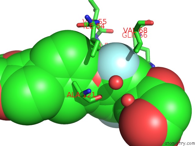
Mono view
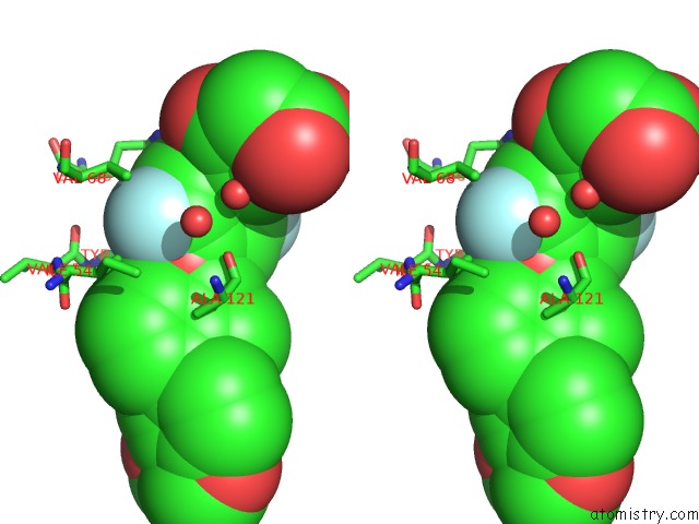
Stereo pair view
Mono view
Stereo pair view
A full contact list of Fluorine with other atoms in the F binding
site number 1 of Structure of Pd-L1/Small-Molecule Inhibitor Complex within 5.0Å range:
|
Fluorine binding site 2 out of 2 in 5n2f
Go back to
Fluorine binding site 2 out
of 2 in the Structure of Pd-L1/Small-Molecule Inhibitor Complex
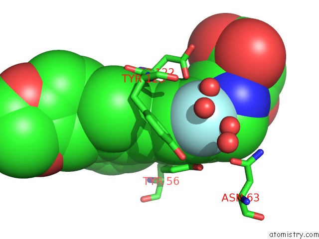
Mono view
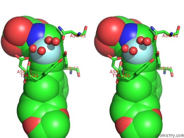
Stereo pair view
Mono view
Stereo pair view
A full contact list of Fluorine with other atoms in the F binding
site number 2 of Structure of Pd-L1/Small-Molecule Inhibitor Complex within 5.0Å range:
|
Reference:
K.Guzik,
K.M.Zak,
P.Grudnik,
K.Magiera,
B.Musielak,
R.Torner,
L.Skalniak,
A.Domling,
G.Dubin,
T.A.Holak.
Small-Molecule Inhibitors of the Programmed Cell Death-1/Programmed Death-Ligand 1 (Pd-1/Pd-L1) Interaction Via Transiently Induced Protein States and Dimerization of Pd-L1. J. Med. Chem. V. 60 5857 2017.
ISSN: ISSN 1520-4804
PubMed: 28613862
DOI: 10.1021/ACS.JMEDCHEM.7B00293
Page generated: Thu Aug 1 11:54:40 2024
ISSN: ISSN 1520-4804
PubMed: 28613862
DOI: 10.1021/ACS.JMEDCHEM.7B00293
Last articles
Zn in 9J0NZn in 9J0O
Zn in 9J0P
Zn in 9FJX
Zn in 9EKB
Zn in 9C0F
Zn in 9CAH
Zn in 9CH0
Zn in 9CH3
Zn in 9CH1