Fluorine »
PDB 5o6j-5olv »
5o9a »
Fluorine in PDB 5o9a: Crystal Structure of the GLUA2 Ligand-Binding Domain (S1S2J-L504Y- N775S) in Complex with Glutamate and BPAM121 at 1.78 A Resolution
Protein crystallography data
The structure of Crystal Structure of the GLUA2 Ligand-Binding Domain (S1S2J-L504Y- N775S) in Complex with Glutamate and BPAM121 at 1.78 A Resolution, PDB code: 5o9a
was solved by
S.Laulumaa,
K.Rovinskaja,
K.A.Frydenvang,
J.S.Kastrup,
with X-Ray Crystallography technique. A brief refinement statistics is given in the table below:
| Resolution Low / High (Å) | 49.20 / 1.78 |
| Space group | P 1 21 1 |
| Cell size a, b, c (Å), α, β, γ (°) | 47.308, 98.408, 121.787, 90.00, 90.53, 90.00 |
| R / Rfree (%) | 15.3 / 17.9 |
Other elements in 5o9a:
The structure of Crystal Structure of the GLUA2 Ligand-Binding Domain (S1S2J-L504Y- N775S) in Complex with Glutamate and BPAM121 at 1.78 A Resolution also contains other interesting chemical elements:
| Chlorine | (Cl) | 11 atoms |
Fluorine Binding Sites:
The binding sites of Fluorine atom in the Crystal Structure of the GLUA2 Ligand-Binding Domain (S1S2J-L504Y- N775S) in Complex with Glutamate and BPAM121 at 1.78 A Resolution
(pdb code 5o9a). This binding sites where shown within
5.0 Angstroms radius around Fluorine atom.
In total 4 binding sites of Fluorine where determined in the Crystal Structure of the GLUA2 Ligand-Binding Domain (S1S2J-L504Y- N775S) in Complex with Glutamate and BPAM121 at 1.78 A Resolution, PDB code: 5o9a:
Jump to Fluorine binding site number: 1; 2; 3; 4;
In total 4 binding sites of Fluorine where determined in the Crystal Structure of the GLUA2 Ligand-Binding Domain (S1S2J-L504Y- N775S) in Complex with Glutamate and BPAM121 at 1.78 A Resolution, PDB code: 5o9a:
Jump to Fluorine binding site number: 1; 2; 3; 4;
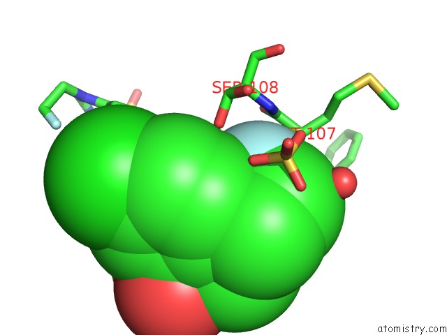
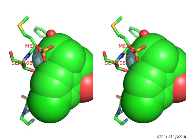
Fluorine binding site 1 out of 4 in 5o9a
Go back to
Fluorine binding site 1 out
of 4 in the Crystal Structure of the GLUA2 Ligand-Binding Domain (S1S2J-L504Y- N775S) in Complex with Glutamate and BPAM121 at 1.78 A Resolution
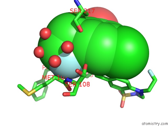
Mono view
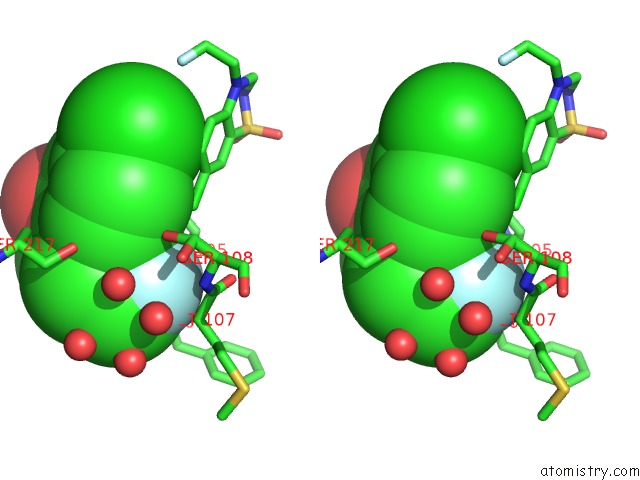
Stereo pair view
Mono view
Stereo pair view
A full contact list of Fluorine with other atoms in the F binding
site number 1 of Crystal Structure of the GLUA2 Ligand-Binding Domain (S1S2J-L504Y- N775S) in Complex with Glutamate and BPAM121 at 1.78 A Resolution within 5.0Å range:
|
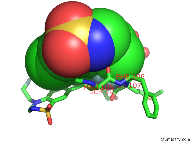
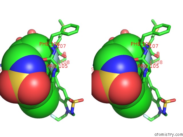
Fluorine binding site 2 out of 4 in 5o9a
Go back to
Fluorine binding site 2 out
of 4 in the Crystal Structure of the GLUA2 Ligand-Binding Domain (S1S2J-L504Y- N775S) in Complex with Glutamate and BPAM121 at 1.78 A Resolution
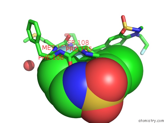
Mono view
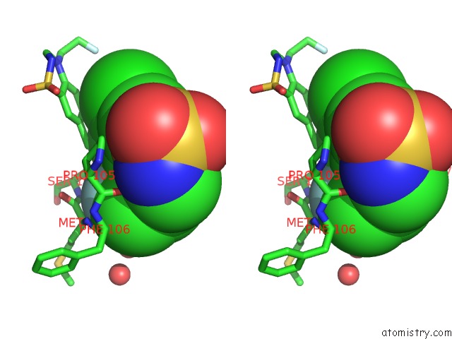
Stereo pair view
Mono view
Stereo pair view
A full contact list of Fluorine with other atoms in the F binding
site number 2 of Crystal Structure of the GLUA2 Ligand-Binding Domain (S1S2J-L504Y- N775S) in Complex with Glutamate and BPAM121 at 1.78 A Resolution within 5.0Å range:
|
Fluorine binding site 3 out of 4 in 5o9a
Go back to
Fluorine binding site 3 out
of 4 in the Crystal Structure of the GLUA2 Ligand-Binding Domain (S1S2J-L504Y- N775S) in Complex with Glutamate and BPAM121 at 1.78 A Resolution
Mono view
Stereo pair view
Mono view
Stereo pair view
A full contact list of Fluorine with other atoms in the F binding
site number 3 of Crystal Structure of the GLUA2 Ligand-Binding Domain (S1S2J-L504Y- N775S) in Complex with Glutamate and BPAM121 at 1.78 A Resolution within 5.0Å range:
|
Fluorine binding site 4 out of 4 in 5o9a
Go back to
Fluorine binding site 4 out
of 4 in the Crystal Structure of the GLUA2 Ligand-Binding Domain (S1S2J-L504Y- N775S) in Complex with Glutamate and BPAM121 at 1.78 A Resolution
Mono view
Stereo pair view
Mono view
Stereo pair view
A full contact list of Fluorine with other atoms in the F binding
site number 4 of Crystal Structure of the GLUA2 Ligand-Binding Domain (S1S2J-L504Y- N775S) in Complex with Glutamate and BPAM121 at 1.78 A Resolution within 5.0Å range:
|
Reference:
E.Goffin,
T.Drapier,
A.P.Larsen,
P.Geubelle,
C.P.Ptak,
S.Laulumaa,
K.Rovinskaja,
J.Gilissen,
P.Tullio,
L.Olsen,
K.Frydenvang,
B.Pirotte,
J.Hanson,
R.E.Oswald,
J.S.Kastrup,
P.Francotte.
7-Phenoxy-Substituted 3,4-Dihydro-2H-1,2,4-Benzothiadiazine 1,1-Dioxides As Positive Allosteric Modulators of Alpha-Amino-3-Hydroxy-5-Methyl-4-Isoxazolepropionic Acid (Ampa) Receptors with Nanomolar Potency. J. Med. Chem. V. 61 251 2018.
ISSN: ISSN 1520-4804
PubMed: 29256599
DOI: 10.1021/ACS.JMEDCHEM.7B01323
Page generated: Thu Aug 1 12:16:40 2024
ISSN: ISSN 1520-4804
PubMed: 29256599
DOI: 10.1021/ACS.JMEDCHEM.7B01323
Last articles
Zn in 9MJ5Zn in 9HNW
Zn in 9G0L
Zn in 9FNE
Zn in 9DZN
Zn in 9E0I
Zn in 9D32
Zn in 9DAK
Zn in 8ZXC
Zn in 8ZUF