Fluorine »
PDB 5o6j-5olv »
5ok2 »
Fluorine in PDB 5ok2: Structure of the D10N Mutant of Beta-Phosphoglucomutase From Lactococcus Lactis Inhibited with Glucose 6-Phosphate and Tetrafluoroaluminate to 1.1A Resolution.
Enzymatic activity of Structure of the D10N Mutant of Beta-Phosphoglucomutase From Lactococcus Lactis Inhibited with Glucose 6-Phosphate and Tetrafluoroaluminate to 1.1A Resolution.
All present enzymatic activity of Structure of the D10N Mutant of Beta-Phosphoglucomutase From Lactococcus Lactis Inhibited with Glucose 6-Phosphate and Tetrafluoroaluminate to 1.1A Resolution.:
5.4.2.6;
5.4.2.6;
Protein crystallography data
The structure of Structure of the D10N Mutant of Beta-Phosphoglucomutase From Lactococcus Lactis Inhibited with Glucose 6-Phosphate and Tetrafluoroaluminate to 1.1A Resolution., PDB code: 5ok2
was solved by
A.J.Robertson,
C.Bisson,
with X-Ray Crystallography technique. A brief refinement statistics is given in the table below:
| Resolution Low / High (Å) | 52.39 / 1.10 |
| Space group | P 21 21 21 |
| Cell size a, b, c (Å), α, β, γ (°) | 37.540, 54.280, 104.670, 90.00, 90.00, 90.00 |
| R / Rfree (%) | 14.8 / 17 |
Other elements in 5ok2:
The structure of Structure of the D10N Mutant of Beta-Phosphoglucomutase From Lactococcus Lactis Inhibited with Glucose 6-Phosphate and Tetrafluoroaluminate to 1.1A Resolution. also contains other interesting chemical elements:
| Magnesium | (Mg) | 2 atoms |
| Aluminium | (Al) | 1 atom |
| Sodium | (Na) | 1 atom |
Fluorine Binding Sites:
The binding sites of Fluorine atom in the Structure of the D10N Mutant of Beta-Phosphoglucomutase From Lactococcus Lactis Inhibited with Glucose 6-Phosphate and Tetrafluoroaluminate to 1.1A Resolution.
(pdb code 5ok2). This binding sites where shown within
5.0 Angstroms radius around Fluorine atom.
In total 4 binding sites of Fluorine where determined in the Structure of the D10N Mutant of Beta-Phosphoglucomutase From Lactococcus Lactis Inhibited with Glucose 6-Phosphate and Tetrafluoroaluminate to 1.1A Resolution., PDB code: 5ok2:
Jump to Fluorine binding site number: 1; 2; 3; 4;
In total 4 binding sites of Fluorine where determined in the Structure of the D10N Mutant of Beta-Phosphoglucomutase From Lactococcus Lactis Inhibited with Glucose 6-Phosphate and Tetrafluoroaluminate to 1.1A Resolution., PDB code: 5ok2:
Jump to Fluorine binding site number: 1; 2; 3; 4;
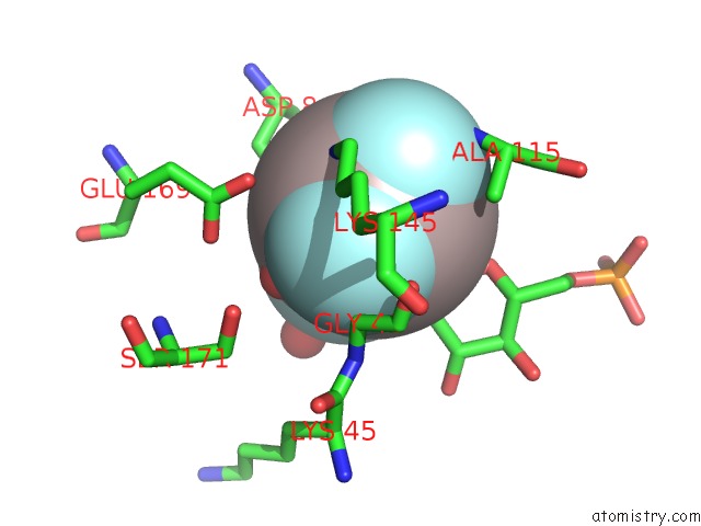
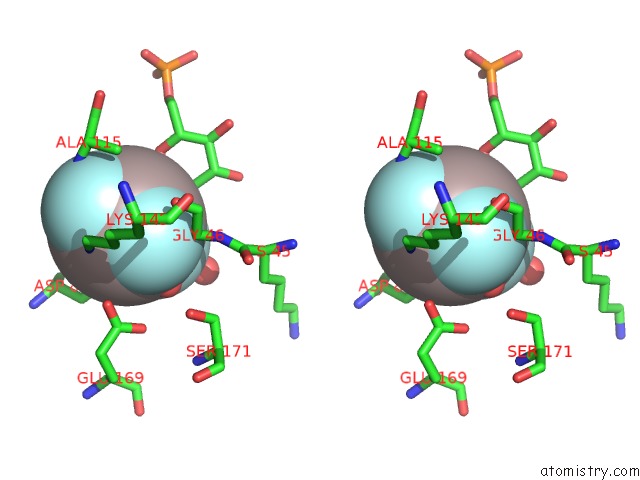
Fluorine binding site 1 out of 4 in 5ok2
Go back to
Fluorine binding site 1 out
of 4 in the Structure of the D10N Mutant of Beta-Phosphoglucomutase From Lactococcus Lactis Inhibited with Glucose 6-Phosphate and Tetrafluoroaluminate to 1.1A Resolution.

Mono view

Stereo pair view
Mono view
Stereo pair view
A full contact list of Fluorine with other atoms in the F binding
site number 1 of Structure of the D10N Mutant of Beta-Phosphoglucomutase From Lactococcus Lactis Inhibited with Glucose 6-Phosphate and Tetrafluoroaluminate to 1.1A Resolution. within 5.0Å range:
|
Fluorine binding site 2 out of 4 in 5ok2
Go back to
Fluorine binding site 2 out
of 4 in the Structure of the D10N Mutant of Beta-Phosphoglucomutase From Lactococcus Lactis Inhibited with Glucose 6-Phosphate and Tetrafluoroaluminate to 1.1A Resolution.
Mono view
Stereo pair view
Mono view
Stereo pair view
A full contact list of Fluorine with other atoms in the F binding
site number 2 of Structure of the D10N Mutant of Beta-Phosphoglucomutase From Lactococcus Lactis Inhibited with Glucose 6-Phosphate and Tetrafluoroaluminate to 1.1A Resolution. within 5.0Å range:
|
Fluorine binding site 3 out of 4 in 5ok2
Go back to
Fluorine binding site 3 out
of 4 in the Structure of the D10N Mutant of Beta-Phosphoglucomutase From Lactococcus Lactis Inhibited with Glucose 6-Phosphate and Tetrafluoroaluminate to 1.1A Resolution.

Mono view
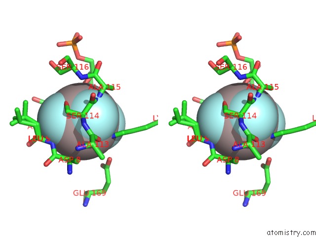
Stereo pair view
Mono view
Stereo pair view
A full contact list of Fluorine with other atoms in the F binding
site number 3 of Structure of the D10N Mutant of Beta-Phosphoglucomutase From Lactococcus Lactis Inhibited with Glucose 6-Phosphate and Tetrafluoroaluminate to 1.1A Resolution. within 5.0Å range:
|
Fluorine binding site 4 out of 4 in 5ok2
Go back to
Fluorine binding site 4 out
of 4 in the Structure of the D10N Mutant of Beta-Phosphoglucomutase From Lactococcus Lactis Inhibited with Glucose 6-Phosphate and Tetrafluoroaluminate to 1.1A Resolution.

Mono view
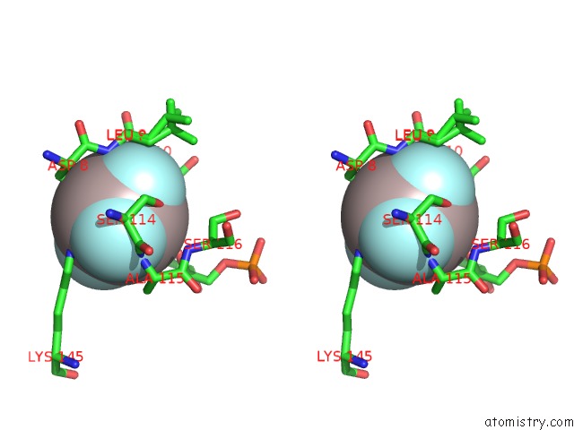
Stereo pair view
Mono view
Stereo pair view
A full contact list of Fluorine with other atoms in the F binding
site number 4 of Structure of the D10N Mutant of Beta-Phosphoglucomutase From Lactococcus Lactis Inhibited with Glucose 6-Phosphate and Tetrafluoroaluminate to 1.1A Resolution. within 5.0Å range:
|
Reference:
L.A.Johnson,
A.J.Robertson,
N.J.Baxter,
C.R.Trevitt,
C.Bisson,
Y.Jin,
H.P.Wood,
A.M.Hounslow,
M.J.Cliff,
G.M.Blackburn,
M.W.Bowler,
J.P.Waltho.
Van Der Waals Contact Between Nucleophile and Transferring Phosphorus Is Insufficient to Achieve Enzyme Transition-State Architecture Acs Catalysis 2018.
ISSN: ESSN 2155-5435
DOI: 10.1021/ACSCATAL.8B01612
Page generated: Thu Aug 1 12:28:53 2024
ISSN: ESSN 2155-5435
DOI: 10.1021/ACSCATAL.8B01612
Last articles
Zn in 9J0NZn in 9J0O
Zn in 9J0P
Zn in 9FJX
Zn in 9EKB
Zn in 9C0F
Zn in 9CAH
Zn in 9CH0
Zn in 9CH3
Zn in 9CH1