Fluorine »
PDB 5qtr-5rah »
5r23 »
Fluorine in PDB 5r23: Pandda Analysis Group Deposition -- Endothiapepsin in Complex with Fragment F2X-Entry F03, Dmso-Free
Enzymatic activity of Pandda Analysis Group Deposition -- Endothiapepsin in Complex with Fragment F2X-Entry F03, Dmso-Free
All present enzymatic activity of Pandda Analysis Group Deposition -- Endothiapepsin in Complex with Fragment F2X-Entry F03, Dmso-Free:
3.4.23.22;
3.4.23.22;
Protein crystallography data
The structure of Pandda Analysis Group Deposition -- Endothiapepsin in Complex with Fragment F2X-Entry F03, Dmso-Free, PDB code: 5r23
was solved by
J.Wollenhaupt,
A.Metz,
T.Barthel,
G.M.A.Lima,
A.Heine,
U.Mueller,
G.Klebe,
M.S.Weiss,
with X-Ray Crystallography technique. A brief refinement statistics is given in the table below:
| Resolution Low / High (Å) | 49.59 / 1.03 |
| Space group | P 1 21 1 |
| Cell size a, b, c (Å), α, β, γ (°) | 45.224, 73.085, 52.586, 90.00, 109.44, 90.00 |
| R / Rfree (%) | 14.6 / 16.6 |
Fluorine Binding Sites:
The binding sites of Fluorine atom in the Pandda Analysis Group Deposition -- Endothiapepsin in Complex with Fragment F2X-Entry F03, Dmso-Free
(pdb code 5r23). This binding sites where shown within
5.0 Angstroms radius around Fluorine atom.
In total only one binding site of Fluorine was determined in the Pandda Analysis Group Deposition -- Endothiapepsin in Complex with Fragment F2X-Entry F03, Dmso-Free, PDB code: 5r23:
In total only one binding site of Fluorine was determined in the Pandda Analysis Group Deposition -- Endothiapepsin in Complex with Fragment F2X-Entry F03, Dmso-Free, PDB code: 5r23:
Fluorine binding site 1 out of 1 in 5r23
Go back to
Fluorine binding site 1 out
of 1 in the Pandda Analysis Group Deposition -- Endothiapepsin in Complex with Fragment F2X-Entry F03, Dmso-Free
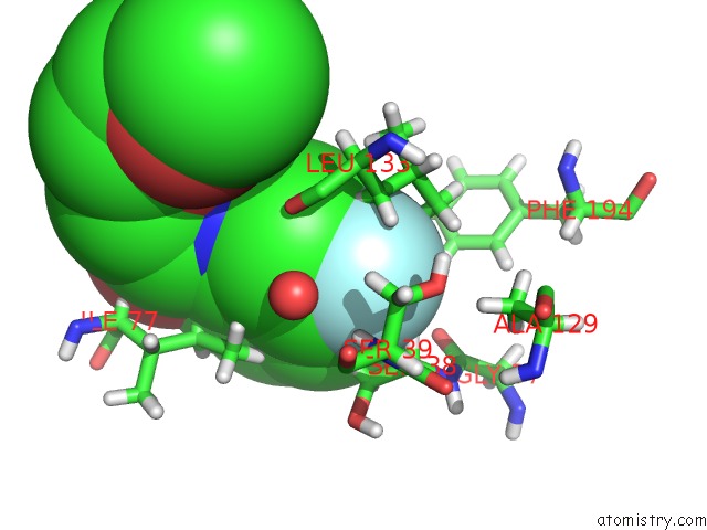
Mono view
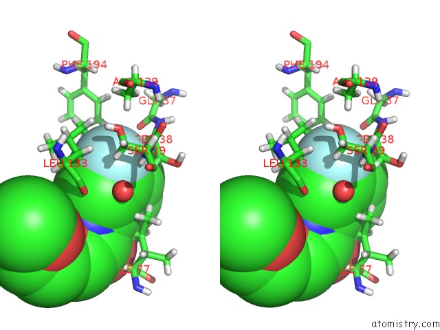
Stereo pair view
Mono view
Stereo pair view
A full contact list of Fluorine with other atoms in the F binding
site number 1 of Pandda Analysis Group Deposition -- Endothiapepsin in Complex with Fragment F2X-Entry F03, Dmso-Free within 5.0Å range:
|
Reference:
J.Wollenhaupt,
A.Metz,
T.Barthel,
G.M.A.Lima,
A.Heine,
U.Mueller,
G.Klebe,
M.S.Weiss.
F2X-Universal and F2X-Entry: Structurally Diverse Compound Libraries For Crystallographic Fragment Screening. Structure 2020.
ISSN: ISSN 0969-2126
PubMed: 32413289
DOI: 10.1016/J.STR.2020.04.019
Page generated: Thu Aug 1 13:33:56 2024
ISSN: ISSN 0969-2126
PubMed: 32413289
DOI: 10.1016/J.STR.2020.04.019
Last articles
Zn in 9J0NZn in 9J0O
Zn in 9J0P
Zn in 9FJX
Zn in 9EKB
Zn in 9C0F
Zn in 9CAH
Zn in 9CH0
Zn in 9CH3
Zn in 9CH1