Fluorine »
PDB 5srx-5tbe »
5swn »
Fluorine in PDB 5swn: Crystal Structure of the Fluoroacetate Dehalogenase RPA1163 - ASP110ASN/Fluoroacetate - Cocrystallized
Enzymatic activity of Crystal Structure of the Fluoroacetate Dehalogenase RPA1163 - ASP110ASN/Fluoroacetate - Cocrystallized
All present enzymatic activity of Crystal Structure of the Fluoroacetate Dehalogenase RPA1163 - ASP110ASN/Fluoroacetate - Cocrystallized:
3.8.1.3;
3.8.1.3;
Protein crystallography data
The structure of Crystal Structure of the Fluoroacetate Dehalogenase RPA1163 - ASP110ASN/Fluoroacetate - Cocrystallized, PDB code: 5swn
was solved by
P.Mehrabi,
T.H.Kim,
S.R.Prosser,
E.F.Pai,
with X-Ray Crystallography technique. A brief refinement statistics is given in the table below:
| Resolution Low / High (Å) | 40.78 / 1.54 |
| Space group | P 1 21 1 |
| Cell size a, b, c (Å), α, β, γ (°) | 41.880, 78.870, 84.900, 90.00, 103.18, 90.00 |
| R / Rfree (%) | 16.9 / 19.4 |
Other elements in 5swn:
The structure of Crystal Structure of the Fluoroacetate Dehalogenase RPA1163 - ASP110ASN/Fluoroacetate - Cocrystallized also contains other interesting chemical elements:
| Chlorine | (Cl) | 2 atoms |
Fluorine Binding Sites:
The binding sites of Fluorine atom in the Crystal Structure of the Fluoroacetate Dehalogenase RPA1163 - ASP110ASN/Fluoroacetate - Cocrystallized
(pdb code 5swn). This binding sites where shown within
5.0 Angstroms radius around Fluorine atom.
In total only one binding site of Fluorine was determined in the Crystal Structure of the Fluoroacetate Dehalogenase RPA1163 - ASP110ASN/Fluoroacetate - Cocrystallized, PDB code: 5swn:
In total only one binding site of Fluorine was determined in the Crystal Structure of the Fluoroacetate Dehalogenase RPA1163 - ASP110ASN/Fluoroacetate - Cocrystallized, PDB code: 5swn:
Fluorine binding site 1 out of 1 in 5swn
Go back to
Fluorine binding site 1 out
of 1 in the Crystal Structure of the Fluoroacetate Dehalogenase RPA1163 - ASP110ASN/Fluoroacetate - Cocrystallized
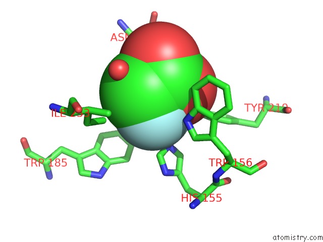
Mono view
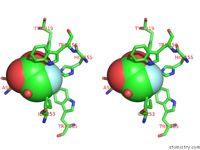
Stereo pair view
Mono view
Stereo pair view
A full contact list of Fluorine with other atoms in the F binding
site number 1 of Crystal Structure of the Fluoroacetate Dehalogenase RPA1163 - ASP110ASN/Fluoroacetate - Cocrystallized within 5.0Å range:
|
Reference:
T.H.Kim,
P.Mehrabi,
Z.Ren,
A.Sljoka,
C.Ing,
A.Bezginov,
L.Ye,
R.Pomes,
R.S.Prosser,
E.F.Pai.
The Role of Dimer Asymmetry and Protomer Dynamics in Enzyme Catalysis. Science V. 355 2017.
ISSN: ESSN 1095-9203
PubMed: 28104837
DOI: 10.1126/SCIENCE.AAG2355
Page generated: Thu Aug 1 14:58:14 2024
ISSN: ESSN 1095-9203
PubMed: 28104837
DOI: 10.1126/SCIENCE.AAG2355
Last articles
Zn in 9JYWZn in 9IR4
Zn in 9IR3
Zn in 9GMX
Zn in 9GMW
Zn in 9JEJ
Zn in 9ERF
Zn in 9ERE
Zn in 9EGV
Zn in 9EGW