Fluorine »
PDB 5sry-5tbm »
5sxc »
Fluorine in PDB 5sxc: Crystal Structure of PI3KALPHA in Complex with Fragment 8
Enzymatic activity of Crystal Structure of PI3KALPHA in Complex with Fragment 8
All present enzymatic activity of Crystal Structure of PI3KALPHA in Complex with Fragment 8:
2.7.1.153; 2.7.11.1;
2.7.1.153; 2.7.11.1;
Protein crystallography data
The structure of Crystal Structure of PI3KALPHA in Complex with Fragment 8, PDB code: 5sxc
was solved by
S.B.Gabelli,
B.Vogelstein,
M.S.Miller,
L.M.Amzel,
with X-Ray Crystallography technique. A brief refinement statistics is given in the table below:
| Resolution Low / High (Å) | 92.46 / 3.55 |
| Space group | P 21 21 21 |
| Cell size a, b, c (Å), α, β, γ (°) | 114.735, 117.271, 150.299, 90.00, 90.00, 90.00 |
| R / Rfree (%) | 19.4 / 26.8 |
Fluorine Binding Sites:
The binding sites of Fluorine atom in the Crystal Structure of PI3KALPHA in Complex with Fragment 8
(pdb code 5sxc). This binding sites where shown within
5.0 Angstroms radius around Fluorine atom.
In total only one binding site of Fluorine was determined in the Crystal Structure of PI3KALPHA in Complex with Fragment 8, PDB code: 5sxc:
In total only one binding site of Fluorine was determined in the Crystal Structure of PI3KALPHA in Complex with Fragment 8, PDB code: 5sxc:
Fluorine binding site 1 out of 1 in 5sxc
Go back to
Fluorine binding site 1 out
of 1 in the Crystal Structure of PI3KALPHA in Complex with Fragment 8
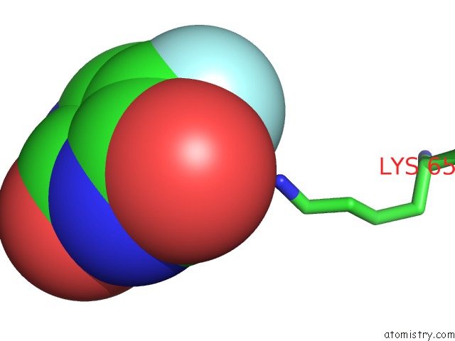
Mono view
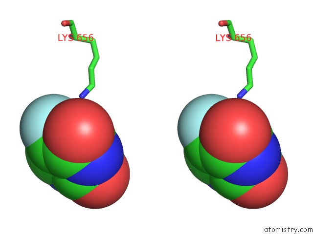
Stereo pair view
Mono view
Stereo pair view
A full contact list of Fluorine with other atoms in the F binding
site number 1 of Crystal Structure of PI3KALPHA in Complex with Fragment 8 within 5.0Å range:
|
Reference:
M.S.Miller,
S.Maheshwari,
F.M.Mcrobb,
K.W.Kinzler,
L.M.Amzel,
B.Vogelstein,
S.B.Gabelli.
Identification of Allosteric Binding Sites For PI3K Alpha Oncogenic Mutant Specific Inhibitor Design. Bioorg. Med. Chem. V. 25 1481 2017.
ISSN: ESSN 1464-3391
PubMed: 28129991
DOI: 10.1016/J.BMC.2017.01.012
Page generated: Tue Jul 15 07:44:37 2025
ISSN: ESSN 1464-3391
PubMed: 28129991
DOI: 10.1016/J.BMC.2017.01.012
Last articles
Fe in 2YXOFe in 2YRS
Fe in 2YXC
Fe in 2YNM
Fe in 2YVJ
Fe in 2YP1
Fe in 2YU2
Fe in 2YU1
Fe in 2YQB
Fe in 2YOO