Fluorine »
PDB 5tbm-5tto »
5te8 »
Fluorine in PDB 5te8: Crystal Structure of the Midazolam-Bound Human CYP3A4
Protein crystallography data
The structure of Crystal Structure of the Midazolam-Bound Human CYP3A4, PDB code: 5te8
was solved by
I.Sevrioukova,
T.Poulos,
with X-Ray Crystallography technique. A brief refinement statistics is given in the table below:
| Resolution Low / High (Å) | 102.34 / 2.70 |
| Space group | P 21 21 21 |
| Cell size a, b, c (Å), α, β, γ (°) | 64.289, 117.980, 205.649, 90.00, 90.00, 90.00 |
| R / Rfree (%) | 22.4 / 29.2 |
Other elements in 5te8:
The structure of Crystal Structure of the Midazolam-Bound Human CYP3A4 also contains other interesting chemical elements:
| Iron | (Fe) | 3 atoms |
| Chlorine | (Cl) | 3 atoms |
Fluorine Binding Sites:
The binding sites of Fluorine atom in the Crystal Structure of the Midazolam-Bound Human CYP3A4
(pdb code 5te8). This binding sites where shown within
5.0 Angstroms radius around Fluorine atom.
In total 3 binding sites of Fluorine where determined in the Crystal Structure of the Midazolam-Bound Human CYP3A4, PDB code: 5te8:
Jump to Fluorine binding site number: 1; 2; 3;
In total 3 binding sites of Fluorine where determined in the Crystal Structure of the Midazolam-Bound Human CYP3A4, PDB code: 5te8:
Jump to Fluorine binding site number: 1; 2; 3;
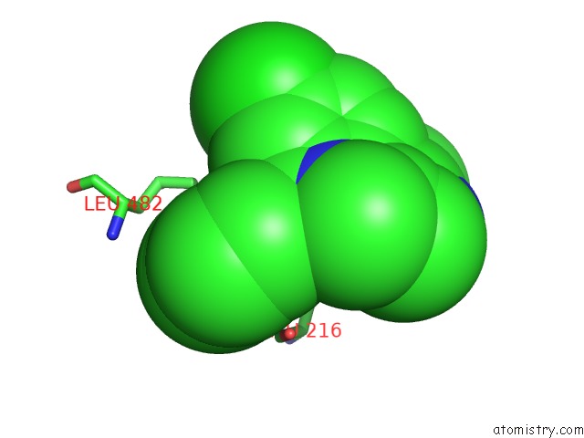
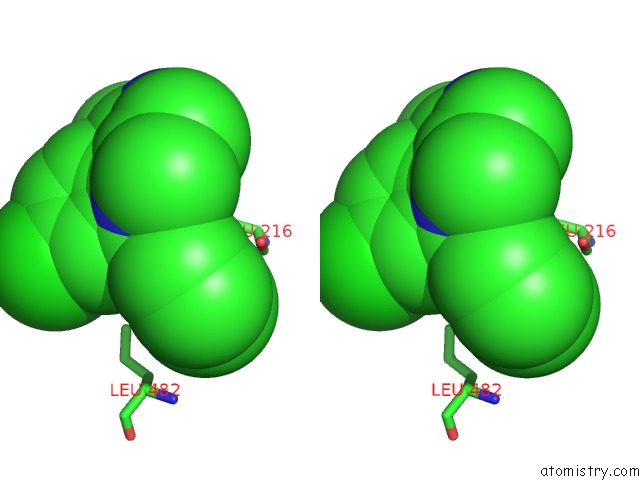
Fluorine binding site 1 out of 3 in 5te8
Go back to
Fluorine binding site 1 out
of 3 in the Crystal Structure of the Midazolam-Bound Human CYP3A4
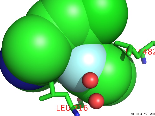
Mono view
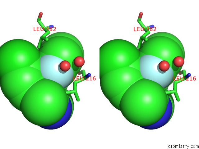
Stereo pair view
Mono view
Stereo pair view
A full contact list of Fluorine with other atoms in the F binding
site number 1 of Crystal Structure of the Midazolam-Bound Human CYP3A4 within 5.0Å range:
|
Fluorine binding site 2 out of 3 in 5te8
Go back to
Fluorine binding site 2 out
of 3 in the Crystal Structure of the Midazolam-Bound Human CYP3A4
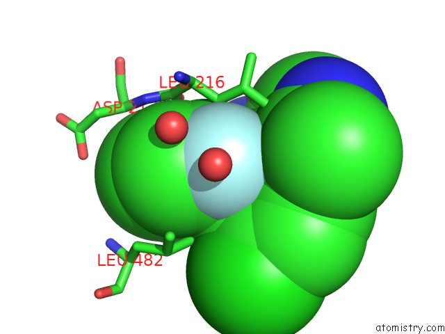
Mono view
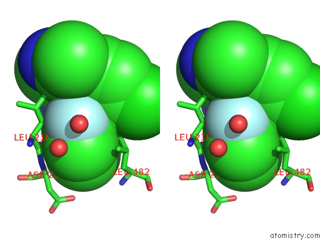
Stereo pair view
Mono view
Stereo pair view
A full contact list of Fluorine with other atoms in the F binding
site number 2 of Crystal Structure of the Midazolam-Bound Human CYP3A4 within 5.0Å range:
|
Fluorine binding site 3 out of 3 in 5te8
Go back to
Fluorine binding site 3 out
of 3 in the Crystal Structure of the Midazolam-Bound Human CYP3A4
Mono view
Stereo pair view
Mono view
Stereo pair view
A full contact list of Fluorine with other atoms in the F binding
site number 3 of Crystal Structure of the Midazolam-Bound Human CYP3A4 within 5.0Å range:
|
Reference:
I.F.Sevrioukova,
T.L.Poulos.
Structural Basis For Regiospecific Midazolam Oxidation By Human Cytochrome P450 3A4. Proc. Natl. Acad. Sci. V. 114 486 2017U.S.A..
ISSN: ESSN 1091-6490
PubMed: 28031486
DOI: 10.1073/PNAS.1616198114
Page generated: Thu Aug 1 15:10:05 2024
ISSN: ESSN 1091-6490
PubMed: 28031486
DOI: 10.1073/PNAS.1616198114
Last articles
Zn in 9J0NZn in 9J0O
Zn in 9J0P
Zn in 9FJX
Zn in 9EKB
Zn in 9C0F
Zn in 9CAH
Zn in 9CH0
Zn in 9CH3
Zn in 9CH1