Fluorine in PDB 5tys: X-Ray Crystal Structure of Wild Type Hiv-1 Protease in Complex with Grl-142
Protein crystallography data
The structure of X-Ray Crystal Structure of Wild Type Hiv-1 Protease in Complex with Grl-142, PDB code: 5tys
was solved by
R.S.Yedidi,
H.Hayashi,
M.Aoki,
D.Das,
A.K.Ghosh,
H.Mitsuya,
with X-Ray Crystallography technique. A brief refinement statistics is given in the table below:
Resolution Low / High (Å)
|
32.86 /
2.01
|
Space group
|
P 61
|
Cell size a, b, c (Å), α, β, γ (°)
|
63.131,
63.131,
82.229,
90.00,
90.00,
120.00
|
R / Rfree (%)
|
19.5 /
23.7
|
Fluorine Binding Sites:
The binding sites of Fluorine atom in the X-Ray Crystal Structure of Wild Type Hiv-1 Protease in Complex with Grl-142
(pdb code 5tys). This binding sites where shown within
5.0 Angstroms radius around Fluorine atom.
In total 4 binding sites of Fluorine where determined in the
X-Ray Crystal Structure of Wild Type Hiv-1 Protease in Complex with Grl-142, PDB code: 5tys:
Jump to Fluorine binding site number:
1;
2;
3;
4;
Fluorine binding site 1 out
of 4 in 5tys
Go back to
Fluorine Binding Sites List in 5tys
Fluorine binding site 1 out
of 4 in the X-Ray Crystal Structure of Wild Type Hiv-1 Protease in Complex with Grl-142
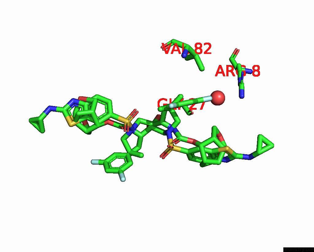
Mono view
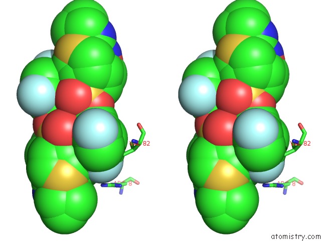
Stereo pair view
|
A full contact list of Fluorine with other atoms in the F binding
site number 1 of X-Ray Crystal Structure of Wild Type Hiv-1 Protease in Complex with Grl-142 within 5.0Å range:
probe
|
atom
|
residue
|
distance (Å)
|
B
|
Occ
|
A:F400
b:23.7
occ:0.47
|
F1
|
A:7OA400
|
0.0
|
23.7
|
0.5
|
C33
|
A:7OA400
|
1.4
|
22.4
|
0.5
|
C06
|
A:7OA400
|
2.4
|
21.2
|
0.5
|
C35
|
A:7OA400
|
2.4
|
20.7
|
0.5
|
C14
|
A:7OA400
|
3.0
|
21.6
|
0.5
|
CG1
|
B:VAL82
|
3.3
|
31.2
|
1.0
|
C11
|
A:7OA400
|
3.3
|
19.2
|
0.5
|
NH2
|
B:ARG8
|
3.5
|
33.3
|
1.0
|
C07
|
A:7OA400
|
3.7
|
20.4
|
0.5
|
C34
|
A:7OA400
|
3.7
|
23.4
|
0.5
|
O
|
B:HOH115
|
3.8
|
26.7
|
1.0
|
CZ
|
B:ARG8
|
4.0
|
40.0
|
1.0
|
C59
|
A:7OA400
|
4.0
|
20.2
|
0.5
|
C12
|
A:7OA400
|
4.1
|
20.3
|
0.5
|
C08
|
A:7OA400
|
4.2
|
20.7
|
0.5
|
C13
|
A:7OA400
|
4.2
|
19.6
|
0.5
|
NH1
|
B:ARG8
|
4.3
|
39.3
|
1.0
|
CB
|
B:VAL82
|
4.5
|
32.8
|
1.0
|
CG2
|
B:VAL82
|
4.6
|
29.5
|
1.0
|
O
|
A:GLY27
|
4.7
|
19.7
|
1.0
|
O2
|
A:7OA400
|
4.7
|
20.4
|
0.5
|
NE
|
B:ARG8
|
4.8
|
32.9
|
1.0
|
C49
|
A:7OA400
|
4.8
|
18.4
|
0.5
|
F2
|
A:7OA400
|
4.8
|
23.7
|
0.5
|
O23
|
A:7OA400
|
4.9
|
17.8
|
0.5
|
C32
|
A:7OA400
|
5.0
|
18.6
|
0.5
|
|
Fluorine binding site 2 out
of 4 in 5tys
Go back to
Fluorine Binding Sites List in 5tys
Fluorine binding site 2 out
of 4 in the X-Ray Crystal Structure of Wild Type Hiv-1 Protease in Complex with Grl-142
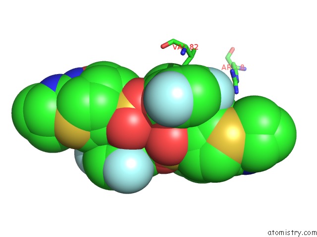
Mono view
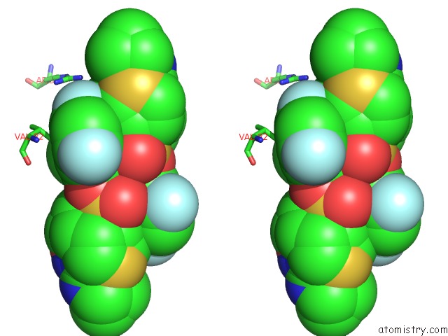
Stereo pair view
|
A full contact list of Fluorine with other atoms in the F binding
site number 2 of X-Ray Crystal Structure of Wild Type Hiv-1 Protease in Complex with Grl-142 within 5.0Å range:
probe
|
atom
|
residue
|
distance (Å)
|
B
|
Occ
|
A:F400
b:24.4
occ:0.53
|
F1
|
A:7OA400
|
0.0
|
24.4
|
0.5
|
C33
|
A:7OA400
|
1.4
|
22.7
|
0.5
|
C06
|
A:7OA400
|
2.4
|
19.1
|
0.5
|
C35
|
A:7OA400
|
2.4
|
21.5
|
0.5
|
C11
|
A:7OA400
|
3.2
|
18.1
|
0.5
|
NH2
|
A:ARG8
|
3.4
|
29.2
|
1.0
|
C07
|
A:7OA400
|
3.7
|
20.4
|
0.5
|
C34
|
A:7OA400
|
3.7
|
22.8
|
0.5
|
C59
|
A:7OA400
|
3.8
|
19.7
|
0.5
|
CZ
|
A:ARG8
|
3.9
|
37.0
|
1.0
|
O
|
B:HOH111
|
3.9
|
25.2
|
1.0
|
NH1
|
A:ARG8
|
3.9
|
40.3
|
1.0
|
CG1
|
A:VAL82
|
3.9
|
29.4
|
1.0
|
C13
|
A:7OA400
|
4.0
|
20.4
|
0.5
|
C12
|
A:7OA400
|
4.0
|
19.9
|
0.5
|
C08
|
A:7OA400
|
4.2
|
20.1
|
0.5
|
CB
|
A:VAL82
|
4.5
|
27.8
|
1.0
|
O2
|
A:7OA400
|
4.6
|
19.6
|
0.5
|
C14
|
A:7OA400
|
4.6
|
21.1
|
0.5
|
C49
|
A:7OA400
|
4.7
|
18.4
|
0.5
|
O23
|
A:7OA400
|
4.8
|
18.0
|
0.5
|
O
|
B:GLY27
|
4.8
|
19.0
|
1.0
|
F2
|
A:7OA400
|
4.8
|
23.1
|
0.5
|
NE
|
A:ARG8
|
4.9
|
34.0
|
1.0
|
C6
|
A:7OA400
|
4.9
|
18.4
|
0.5
|
C32
|
A:7OA400
|
5.0
|
18.6
|
0.5
|
CG2
|
A:VAL82
|
5.0
|
26.4
|
1.0
|
|
Fluorine binding site 3 out
of 4 in 5tys
Go back to
Fluorine Binding Sites List in 5tys
Fluorine binding site 3 out
of 4 in the X-Ray Crystal Structure of Wild Type Hiv-1 Protease in Complex with Grl-142
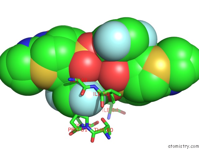
Mono view
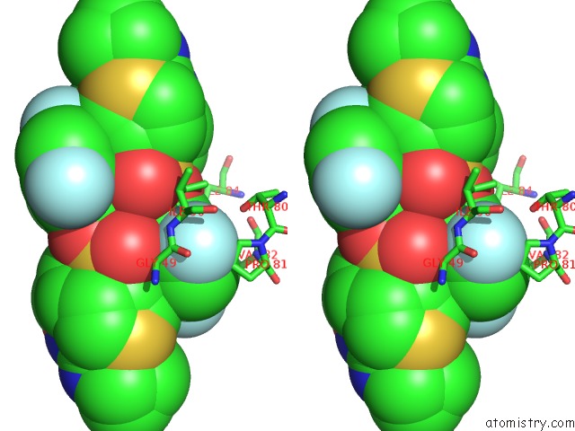
Stereo pair view
|
A full contact list of Fluorine with other atoms in the F binding
site number 3 of X-Ray Crystal Structure of Wild Type Hiv-1 Protease in Complex with Grl-142 within 5.0Å range:
probe
|
atom
|
residue
|
distance (Å)
|
B
|
Occ
|
A:F400
b:23.7
occ:0.47
|
F2
|
A:7OA400
|
0.0
|
23.7
|
0.5
|
C34
|
A:7OA400
|
1.4
|
23.4
|
0.5
|
C08
|
A:7OA400
|
2.4
|
20.7
|
0.5
|
C35
|
A:7OA400
|
2.4
|
20.7
|
0.5
|
CG
|
B:PRO81
|
2.8
|
29.8
|
1.0
|
N
|
A:ILE50
|
3.0
|
21.2
|
1.0
|
C
|
A:GLY49
|
3.0
|
24.3
|
1.0
|
CD
|
B:PRO81
|
3.1
|
28.9
|
1.0
|
C15
|
A:7OA400
|
3.1
|
20.4
|
0.5
|
CA
|
A:ILE50
|
3.4
|
25.7
|
1.0
|
CA
|
A:GLY49
|
3.5
|
24.0
|
1.0
|
O
|
A:GLY49
|
3.5
|
21.1
|
1.0
|
C07
|
A:7OA400
|
3.6
|
20.4
|
0.5
|
C33
|
A:7OA400
|
3.7
|
22.4
|
0.5
|
CG1
|
A:ILE50
|
3.8
|
32.8
|
1.0
|
C14
|
A:7OA400
|
4.0
|
21.6
|
0.5
|
C13
|
A:7OA400
|
4.1
|
19.6
|
0.5
|
CD1
|
A:ILE50
|
4.1
|
30.5
|
1.0
|
C06
|
A:7OA400
|
4.2
|
21.2
|
0.5
|
CB
|
A:ILE50
|
4.2
|
26.0
|
1.0
|
CB
|
B:PRO81
|
4.2
|
28.2
|
1.0
|
N
|
B:PRO81
|
4.3
|
29.9
|
1.0
|
OG1
|
B:THR80
|
4.3
|
23.2
|
1.0
|
O
|
A:HOH503
|
4.5
|
20.5
|
1.0
|
N
|
A:GLY49
|
4.5
|
25.0
|
1.0
|
C12
|
A:7OA400
|
4.5
|
20.3
|
0.5
|
C
|
A:ILE50
|
4.6
|
27.9
|
1.0
|
O10
|
A:7OA400
|
4.6
|
17.9
|
0.5
|
O9
|
A:7OA400
|
4.7
|
20.9
|
0.5
|
O22
|
A:7OA400
|
4.8
|
18.0
|
0.5
|
F1
|
A:7OA400
|
4.8
|
23.7
|
0.5
|
CA
|
B:PRO81
|
4.9
|
31.3
|
1.0
|
C32
|
A:7OA400
|
4.9
|
18.6
|
0.5
|
N
|
B:VAL82
|
4.9
|
28.9
|
1.0
|
CG1
|
B:VAL82
|
4.9
|
31.2
|
1.0
|
O
|
A:ILE50
|
4.9
|
33.7
|
1.0
|
CD1
|
B:ILE84
|
5.0
|
26.4
|
1.0
|
|
Fluorine binding site 4 out
of 4 in 5tys
Go back to
Fluorine Binding Sites List in 5tys
Fluorine binding site 4 out
of 4 in the X-Ray Crystal Structure of Wild Type Hiv-1 Protease in Complex with Grl-142
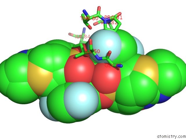
Mono view
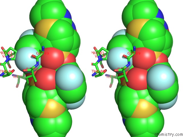
Stereo pair view
|
A full contact list of Fluorine with other atoms in the F binding
site number 4 of X-Ray Crystal Structure of Wild Type Hiv-1 Protease in Complex with Grl-142 within 5.0Å range:
probe
|
atom
|
residue
|
distance (Å)
|
B
|
Occ
|
A:F400
b:23.1
occ:0.53
|
F2
|
A:7OA400
|
0.0
|
23.1
|
0.5
|
C34
|
A:7OA400
|
1.4
|
22.8
|
0.5
|
C08
|
A:7OA400
|
2.4
|
20.1
|
0.5
|
C35
|
A:7OA400
|
2.4
|
21.5
|
0.5
|
CG
|
A:PRO81
|
2.9
|
30.0
|
1.0
|
N
|
B:ILE50
|
3.0
|
22.1
|
1.0
|
CD
|
A:PRO81
|
3.0
|
26.8
|
1.0
|
C
|
B:GLY49
|
3.1
|
26.2
|
1.0
|
CA
|
B:ILE50
|
3.2
|
24.3
|
1.0
|
C15
|
A:7OA400
|
3.4
|
20.3
|
0.5
|
O
|
B:GLY49
|
3.5
|
22.6
|
1.0
|
C07
|
A:7OA400
|
3.6
|
20.4
|
0.5
|
CA
|
B:GLY49
|
3.7
|
24.6
|
1.0
|
C33
|
A:7OA400
|
3.7
|
22.7
|
0.5
|
CB
|
B:ILE50
|
3.8
|
26.7
|
1.0
|
C13
|
A:7OA400
|
3.9
|
20.4
|
0.5
|
CG1
|
B:ILE50
|
3.9
|
25.7
|
1.0
|
C06
|
A:7OA400
|
4.2
|
19.1
|
0.5
|
OG1
|
A:THR80
|
4.2
|
23.9
|
1.0
|
CG1
|
A:VAL82
|
4.3
|
29.4
|
1.0
|
N
|
A:PRO81
|
4.3
|
31.3
|
1.0
|
CB
|
A:PRO81
|
4.3
|
28.0
|
1.0
|
C
|
B:ILE50
|
4.5
|
30.1
|
1.0
|
O9
|
A:7OA400
|
4.6
|
20.3
|
0.5
|
O
|
A:HOH503
|
4.6
|
20.5
|
1.0
|
C12
|
A:7OA400
|
4.7
|
19.9
|
0.5
|
N
|
B:GLY49
|
4.7
|
24.1
|
1.0
|
F1
|
A:7OA400
|
4.8
|
24.4
|
0.5
|
O10
|
A:7OA400
|
4.9
|
19.8
|
0.5
|
O
|
B:ILE50
|
4.9
|
34.4
|
1.0
|
N
|
A:VAL82
|
4.9
|
28.1
|
1.0
|
CD1
|
B:ILE50
|
4.9
|
24.5
|
1.0
|
C32
|
A:7OA400
|
4.9
|
18.6
|
0.5
|
CA
|
A:PRO81
|
4.9
|
32.3
|
1.0
|
CD1
|
A:ILE84
|
5.0
|
25.3
|
1.0
|
|
Reference:
M.Aoki,
H.Hayashi,
K.V.Rao,
D.Das,
N.Higashi-Kuwata,
H.Bulut,
H.Aoki-Ogata,
Y.Takamatsu,
R.S.Yedidi,
D.A.Davis,
S.I.Hattori,
N.Nishida,
K.Hasegawa,
N.Takamune,
P.R.Nyalapatla,
H.L.Osswald,
H.Jono,
H.Saito,
R.Yarchoan,
S.Misumi,
A.K.Ghosh,
H.Mitsuya.
A Novel Central Nervous System-Penetrating Protease Inhibitor Overcomes Human Immunodeficiency Virus 1 Resistance with Unprecedented Am to Pm Potency. Elife V. 6 2017.
ISSN: ESSN 2050-084X
PubMed: 29039736
DOI: 10.7554/ELIFE.28020
Page generated: Thu Aug 1 15:27:10 2024
|