Fluorine »
PDB 5ug9-5uv2 »
5uos »
Fluorine in PDB 5uos: Crystal Structure of Cblc (Mmachc) (1-238), A Human B12 Processing Enzyme, Complexed with An Antivitamin B12
Protein crystallography data
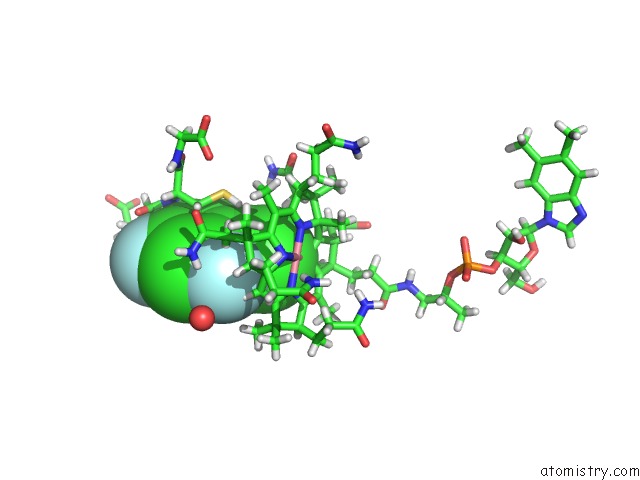
The structure of Crystal Structure of Cblc (Mmachc) (1-238), A Human B12 Processing Enzyme, Complexed with An Antivitamin B12, PDB code: 5uos
was solved by
A.Shanmuganathan,
A.Karasik,
M.Ruetz,
R.Banerjee,
B.Krautler,
M.Koutmos,
with X-Ray Crystallography technique. A brief refinement statistics is given in the table below:
| Resolution Low / High (Å) | 99.10 / 2.51 |
| Space group | P 61 2 2 |
| Cell size a, b, c (Å), α, β, γ (°) | 114.432, 114.432, 150.631, 90.00, 90.00, 120.00 |
| R / Rfree (%) | 17.6 / 21.4 |
Other elements in 5uos:
The structure of Crystal Structure of Cblc (Mmachc) (1-238), A Human B12 Processing Enzyme, Complexed with An Antivitamin B12 also contains other interesting chemical elements:
| Cobalt | (Co) | 1 atom |
| Sodium | (Na) | 1 atom |
Fluorine Binding Sites:
The binding sites of Fluorine atom in the Crystal Structure of Cblc (Mmachc) (1-238), A Human B12 Processing Enzyme, Complexed with An Antivitamin B12
(pdb code 5uos). This binding sites where shown within
5.0 Angstroms radius around Fluorine atom.
In total 2 binding sites of Fluorine where determined in the Crystal Structure of Cblc (Mmachc) (1-238), A Human B12 Processing Enzyme, Complexed with An Antivitamin B12, PDB code: 5uos:
Jump to Fluorine binding site number: 1; 2;
In total 2 binding sites of Fluorine where determined in the Crystal Structure of Cblc (Mmachc) (1-238), A Human B12 Processing Enzyme, Complexed with An Antivitamin B12, PDB code: 5uos:
Jump to Fluorine binding site number: 1; 2;
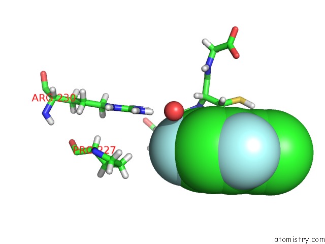
Fluorine binding site 1 out of 2 in 5uos
Go back to
Fluorine binding site 1 out
of 2 in the Crystal Structure of Cblc (Mmachc) (1-238), A Human B12 Processing Enzyme, Complexed with An Antivitamin B12
Mono view
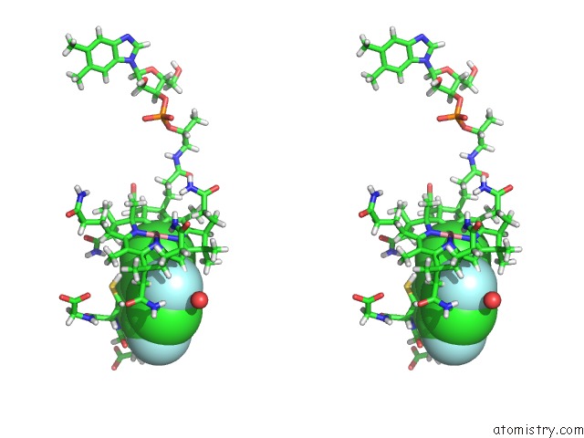
Stereo pair view
Mono view
Stereo pair view
A full contact list of Fluorine with other atoms in the F binding
site number 1 of Crystal Structure of Cblc (Mmachc) (1-238), A Human B12 Processing Enzyme, Complexed with An Antivitamin B12 within 5.0Å range:
|
Fluorine binding site 2 out of 2 in 5uos
Go back to
Fluorine binding site 2 out
of 2 in the Crystal Structure of Cblc (Mmachc) (1-238), A Human B12 Processing Enzyme, Complexed with An Antivitamin B12
Mono view
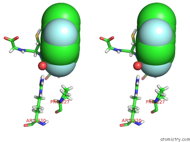
Stereo pair view
Mono view
Stereo pair view
A full contact list of Fluorine with other atoms in the F binding
site number 2 of Crystal Structure of Cblc (Mmachc) (1-238), A Human B12 Processing Enzyme, Complexed with An Antivitamin B12 within 5.0Å range:
|
Reference:
M.Ruetz,
A.Shanmuganathan,
C.Gherasim,
A.Karasik,
R.Salchner,
C.Kieninger,
K.Wurst,
R.Banerjee,
M.Koutmos,
B.Krautler.
Antivitamin B12 Inhibition of the Human B12 -Processing Enzyme Cblc: Crystal Structure of An Inactive Ternary Complex with Glutathione As the Cosubstrate. Angew. Chem. Int. Ed. Engl. V. 56 7387 2017.
ISSN: ESSN 1521-3773
PubMed: 28544088
DOI: 10.1002/ANIE.201701583
Page generated: Tue Jul 15 08:16:29 2025
ISSN: ESSN 1521-3773
PubMed: 28544088
DOI: 10.1002/ANIE.201701583
Last articles
Fe in 2YXOFe in 2YRS
Fe in 2YXC
Fe in 2YNM
Fe in 2YVJ
Fe in 2YP1
Fe in 2YU2
Fe in 2YU1
Fe in 2YQB
Fe in 2YOO