Fluorine »
PDB 5uv3-5vd4 »
5vd2 »
Fluorine in PDB 5vd2: Crystal Structure of Human WEE1 Kinase Domain in Complex with Pf- 03814735
Enzymatic activity of Crystal Structure of Human WEE1 Kinase Domain in Complex with Pf- 03814735
All present enzymatic activity of Crystal Structure of Human WEE1 Kinase Domain in Complex with Pf- 03814735:
2.7.10.2;
2.7.10.2;
Protein crystallography data
The structure of Crystal Structure of Human WEE1 Kinase Domain in Complex with Pf- 03814735, PDB code: 5vd2
was solved by
J.-Y.Zhu,
E.Schonbrunn,
with X-Ray Crystallography technique. A brief refinement statistics is given in the table below:
| Resolution Low / High (Å) | 34.11 / 2.05 |
| Space group | P 1 21 1 |
| Cell size a, b, c (Å), α, β, γ (°) | 50.520, 45.710, 59.440, 90.00, 102.52, 90.00 |
| R / Rfree (%) | 20.3 / 25.6 |
Other elements in 5vd2:
The structure of Crystal Structure of Human WEE1 Kinase Domain in Complex with Pf- 03814735 also contains other interesting chemical elements:
| Chlorine | (Cl) | 2 atoms |
Fluorine Binding Sites:
The binding sites of Fluorine atom in the Crystal Structure of Human WEE1 Kinase Domain in Complex with Pf- 03814735
(pdb code 5vd2). This binding sites where shown within
5.0 Angstroms radius around Fluorine atom.
In total 3 binding sites of Fluorine where determined in the Crystal Structure of Human WEE1 Kinase Domain in Complex with Pf- 03814735, PDB code: 5vd2:
Jump to Fluorine binding site number: 1; 2; 3;
In total 3 binding sites of Fluorine where determined in the Crystal Structure of Human WEE1 Kinase Domain in Complex with Pf- 03814735, PDB code: 5vd2:
Jump to Fluorine binding site number: 1; 2; 3;
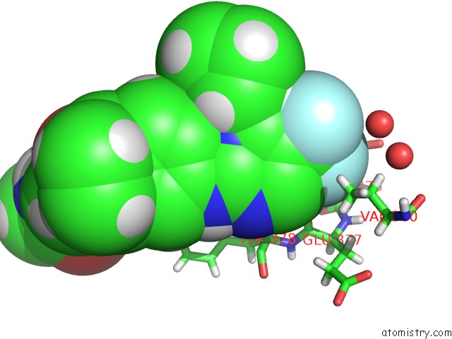
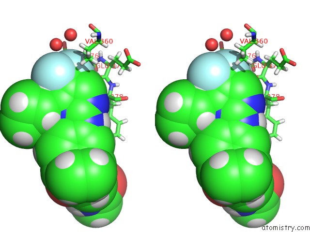
Fluorine binding site 1 out of 3 in 5vd2
Go back to
Fluorine binding site 1 out
of 3 in the Crystal Structure of Human WEE1 Kinase Domain in Complex with Pf- 03814735
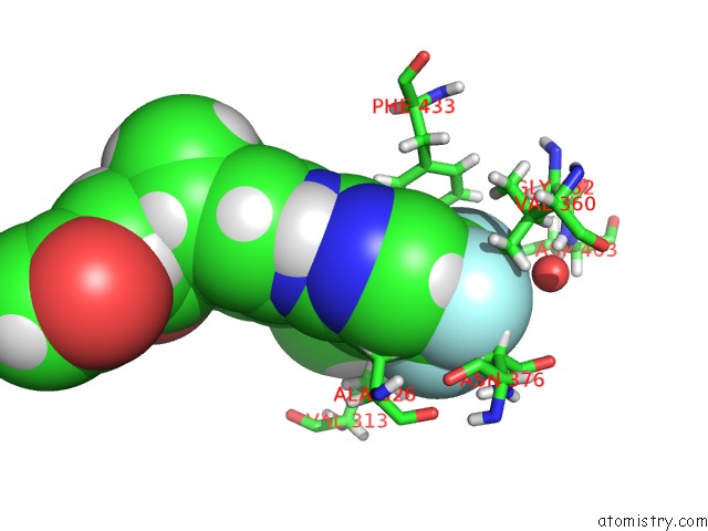
Mono view
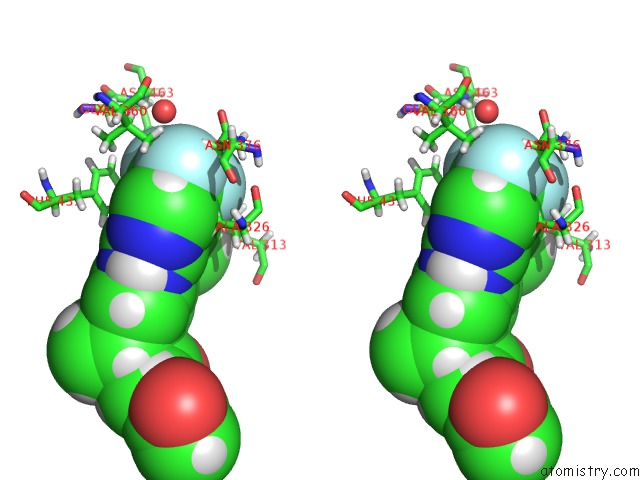
Stereo pair view
Mono view
Stereo pair view
A full contact list of Fluorine with other atoms in the F binding
site number 1 of Crystal Structure of Human WEE1 Kinase Domain in Complex with Pf- 03814735 within 5.0Å range:
|
Fluorine binding site 2 out of 3 in 5vd2
Go back to
Fluorine binding site 2 out
of 3 in the Crystal Structure of Human WEE1 Kinase Domain in Complex with Pf- 03814735
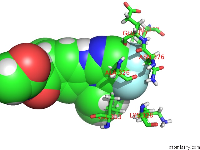
Mono view
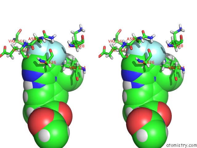
Stereo pair view
Mono view
Stereo pair view
A full contact list of Fluorine with other atoms in the F binding
site number 2 of Crystal Structure of Human WEE1 Kinase Domain in Complex with Pf- 03814735 within 5.0Å range:
|
Fluorine binding site 3 out of 3 in 5vd2
Go back to
Fluorine binding site 3 out
of 3 in the Crystal Structure of Human WEE1 Kinase Domain in Complex with Pf- 03814735
Mono view
Stereo pair view
Mono view
Stereo pair view
A full contact list of Fluorine with other atoms in the F binding
site number 3 of Crystal Structure of Human WEE1 Kinase Domain in Complex with Pf- 03814735 within 5.0Å range:
|
Reference:
J.Y.Zhu,
R.A.Cuellar,
N.Berndt,
H.E.Lee,
S.H.Olesen,
M.P.Martin,
J.T.Jensen,
G.I.Georg,
E.Schonbrunn.
Structural Basis of Wee Kinases Functionality and Inactivation By Diverse Small Molecule Inhibitors. J. Med. Chem. V. 60 7863 2017.
ISSN: ISSN 1520-4804
PubMed: 28792760
DOI: 10.1021/ACS.JMEDCHEM.7B00996
Page generated: Tue Jul 15 08:33:35 2025
ISSN: ISSN 1520-4804
PubMed: 28792760
DOI: 10.1021/ACS.JMEDCHEM.7B00996
Last articles
Fe in 2YXOFe in 2YRS
Fe in 2YXC
Fe in 2YNM
Fe in 2YVJ
Fe in 2YP1
Fe in 2YU2
Fe in 2YU1
Fe in 2YQB
Fe in 2YOO