Fluorine »
PDB 5vta-5wel »
5wbq »
Fluorine in PDB 5wbq: Structure of Human Ketohexokinase Complexed with Hits From Fragment Screening
Enzymatic activity of Structure of Human Ketohexokinase Complexed with Hits From Fragment Screening
All present enzymatic activity of Structure of Human Ketohexokinase Complexed with Hits From Fragment Screening:
2.7.1.3;
2.7.1.3;
Protein crystallography data
The structure of Structure of Human Ketohexokinase Complexed with Hits From Fragment Screening, PDB code: 5wbq
was solved by
J.Pandit,
with X-Ray Crystallography technique. A brief refinement statistics is given in the table below:
| Resolution Low / High (Å) | 34.70 / 2.40 |
| Space group | P 21 21 21 |
| Cell size a, b, c (Å), α, β, γ (°) | 83.200, 85.410, 138.690, 90.00, 90.00, 90.00 |
| R / Rfree (%) | 20.3 / 24.8 |
Other elements in 5wbq:
The structure of Structure of Human Ketohexokinase Complexed with Hits From Fragment Screening also contains other interesting chemical elements:
| Chlorine | (Cl) | 1 atom |
Fluorine Binding Sites:
The binding sites of Fluorine atom in the Structure of Human Ketohexokinase Complexed with Hits From Fragment Screening
(pdb code 5wbq). This binding sites where shown within
5.0 Angstroms radius around Fluorine atom.
In total 6 binding sites of Fluorine where determined in the Structure of Human Ketohexokinase Complexed with Hits From Fragment Screening, PDB code: 5wbq:
Jump to Fluorine binding site number: 1; 2; 3; 4; 5; 6;
In total 6 binding sites of Fluorine where determined in the Structure of Human Ketohexokinase Complexed with Hits From Fragment Screening, PDB code: 5wbq:
Jump to Fluorine binding site number: 1; 2; 3; 4; 5; 6;
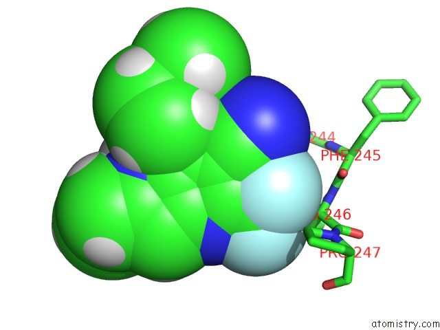
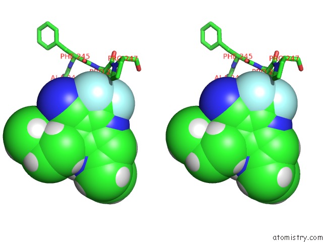
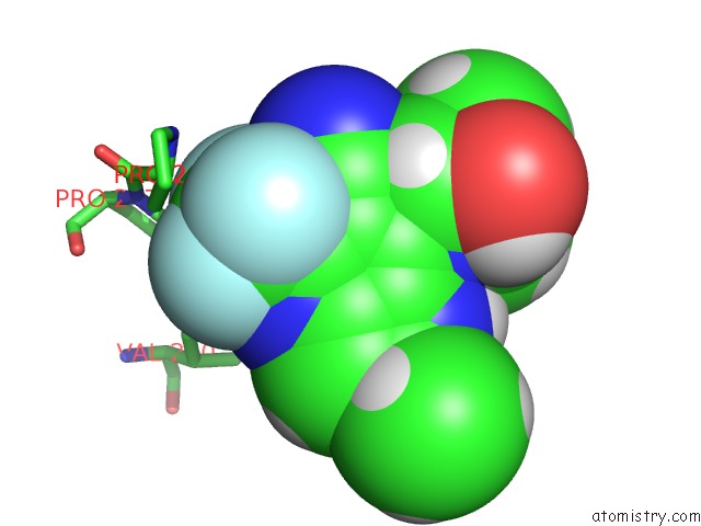
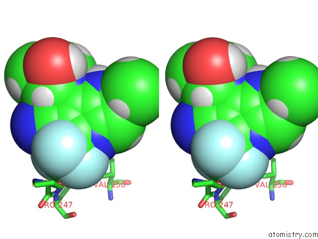
Fluorine binding site 1 out of 6 in 5wbq
Go back to
Fluorine binding site 1 out
of 6 in the Structure of Human Ketohexokinase Complexed with Hits From Fragment Screening
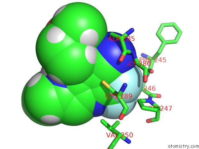
Mono view
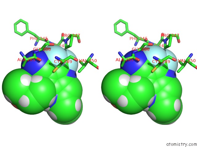
Stereo pair view
Mono view
Stereo pair view
A full contact list of Fluorine with other atoms in the F binding
site number 1 of Structure of Human Ketohexokinase Complexed with Hits From Fragment Screening within 5.0Å range:
|
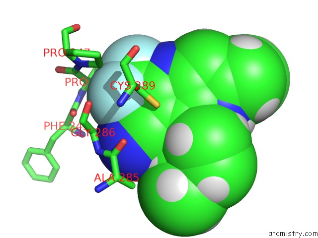
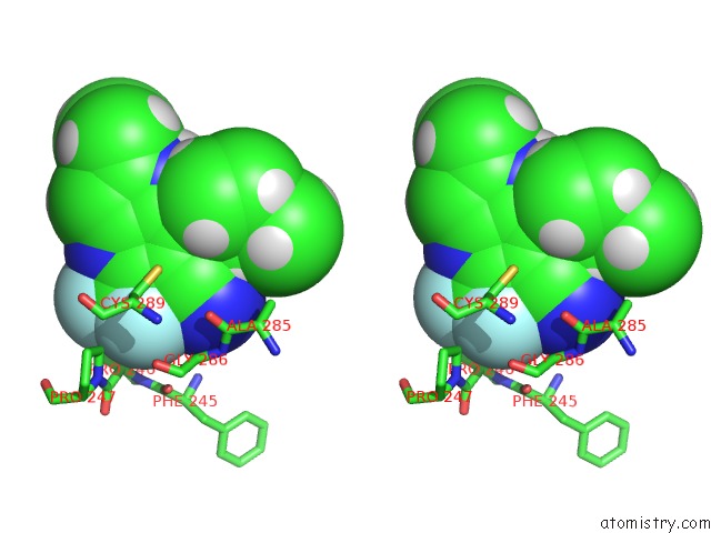
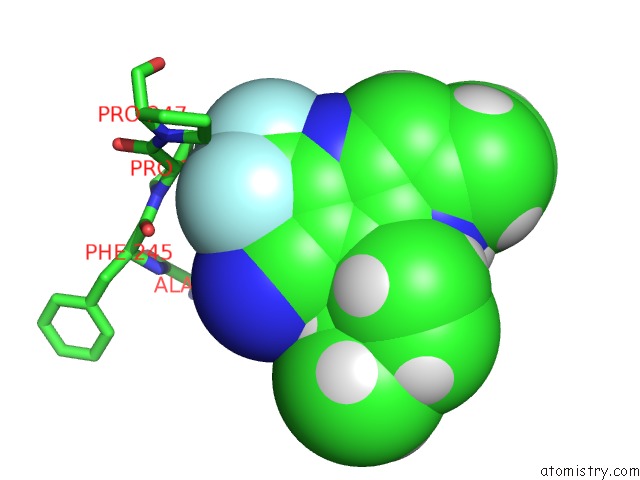
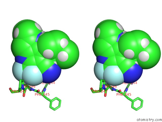
Fluorine binding site 2 out of 6 in 5wbq
Go back to
Fluorine binding site 2 out
of 6 in the Structure of Human Ketohexokinase Complexed with Hits From Fragment Screening
Mono view
Stereo pair view
Mono view
Stereo pair view
A full contact list of Fluorine with other atoms in the F binding
site number 2 of Structure of Human Ketohexokinase Complexed with Hits From Fragment Screening within 5.0Å range:
|
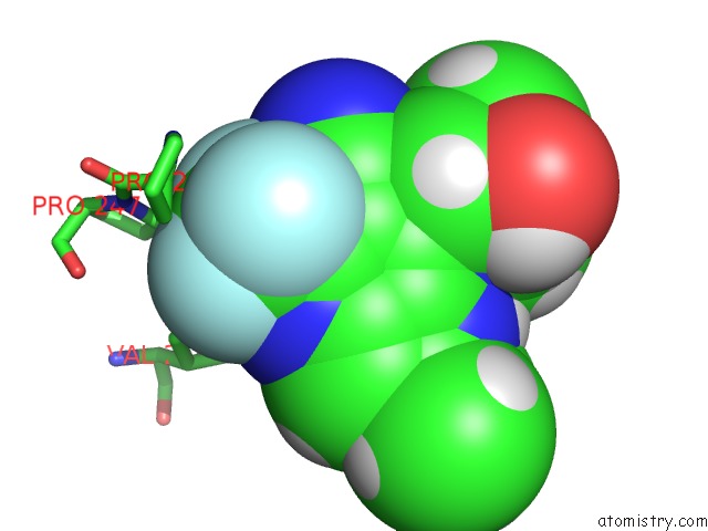
Fluorine binding site 3 out of 6 in 5wbq
Go back to
Fluorine binding site 3 out
of 6 in the Structure of Human Ketohexokinase Complexed with Hits From Fragment Screening
Mono view
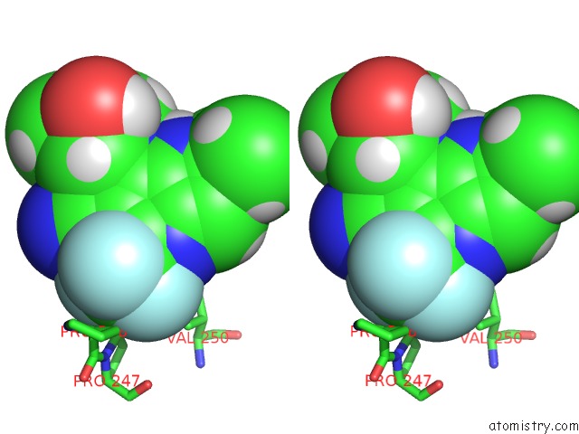
Stereo pair view
Mono view
Stereo pair view
A full contact list of Fluorine with other atoms in the F binding
site number 3 of Structure of Human Ketohexokinase Complexed with Hits From Fragment Screening within 5.0Å range:
|
Fluorine binding site 4 out of 6 in 5wbq
Go back to
Fluorine binding site 4 out
of 6 in the Structure of Human Ketohexokinase Complexed with Hits From Fragment Screening
Mono view
Stereo pair view
Mono view
Stereo pair view
A full contact list of Fluorine with other atoms in the F binding
site number 4 of Structure of Human Ketohexokinase Complexed with Hits From Fragment Screening within 5.0Å range:
|
Fluorine binding site 5 out of 6 in 5wbq
Go back to
Fluorine binding site 5 out
of 6 in the Structure of Human Ketohexokinase Complexed with Hits From Fragment Screening
Mono view
Stereo pair view
Mono view
Stereo pair view
A full contact list of Fluorine with other atoms in the F binding
site number 5 of Structure of Human Ketohexokinase Complexed with Hits From Fragment Screening within 5.0Å range:
|
Fluorine binding site 6 out of 6 in 5wbq
Go back to
Fluorine binding site 6 out
of 6 in the Structure of Human Ketohexokinase Complexed with Hits From Fragment Screening
Mono view
Stereo pair view
Mono view
Stereo pair view
A full contact list of Fluorine with other atoms in the F binding
site number 6 of Structure of Human Ketohexokinase Complexed with Hits From Fragment Screening within 5.0Å range:
|
Reference:
K.Huard,
K.Ahn,
P.Amor,
D.A.Beebe,
K.A.Borzilleri,
B.A.Chrunyk,
S.B.Coffey,
Y.Cong,
E.L.Conn,
J.S.Culp,
M.S.Dowling,
M.F.Gorgoglione,
J.A.Gutierrez,
J.D.Knafels,
E.A.Lachapelle,
J.Pandit,
K.D.Parris,
S.Perez,
J.A.Pfefferkorn,
D.A.Price,
B.Raymer,
T.T.Ross,
A.Shavnya,
A.C.Smith,
T.A.Subashi,
G.J.Tesz,
B.A.Thuma,
M.Tu,
J.D.Weaver,
Y.Weng,
J.M.Withka,
G.Xing,
T.V.Magee.
Discovery of Fragment-Derived Small Molecules For in Vivo Inhibition of Ketohexokinase (Khk). J. Med. Chem. V. 60 7835 2017.
ISSN: ISSN 1520-4804
PubMed: 28853885
DOI: 10.1021/ACS.JMEDCHEM.7B00947
Page generated: Thu Aug 1 16:30:44 2024
ISSN: ISSN 1520-4804
PubMed: 28853885
DOI: 10.1021/ACS.JMEDCHEM.7B00947
Last articles
Zn in 9J0NZn in 9J0O
Zn in 9J0P
Zn in 9FJX
Zn in 9EKB
Zn in 9C0F
Zn in 9CAH
Zn in 9CH0
Zn in 9CH3
Zn in 9CH1