Fluorine »
PDB 5y6d-5zab »
5yak »
Fluorine in PDB 5yak: The Crystal Structure of Human Iyd THR239 Mutant with Ligand 3- Fluorotyrosine (F-Tyr)
Enzymatic activity of The Crystal Structure of Human Iyd THR239 Mutant with Ligand 3- Fluorotyrosine (F-Tyr)
All present enzymatic activity of The Crystal Structure of Human Iyd THR239 Mutant with Ligand 3- Fluorotyrosine (F-Tyr):
1.21.1.1;
1.21.1.1;
Protein crystallography data
The structure of The Crystal Structure of Human Iyd THR239 Mutant with Ligand 3- Fluorotyrosine (F-Tyr), PDB code: 5yak
was solved by
J.M.Hu,
S.E.Rokita,
J.Schlessman,
with X-Ray Crystallography technique. A brief refinement statistics is given in the table below:
| Resolution Low / High (Å) | 46.52 / 2.30 |
| Space group | P 61 |
| Cell size a, b, c (Å), α, β, γ (°) | 105.079, 105.079, 300.202, 90.00, 90.00, 120.00 |
| R / Rfree (%) | 18.3 / 22.6 |
Fluorine Binding Sites:
The binding sites of Fluorine atom in the The Crystal Structure of Human Iyd THR239 Mutant with Ligand 3- Fluorotyrosine (F-Tyr)
(pdb code 5yak). This binding sites where shown within
5.0 Angstroms radius around Fluorine atom.
In total 6 binding sites of Fluorine where determined in the The Crystal Structure of Human Iyd THR239 Mutant with Ligand 3- Fluorotyrosine (F-Tyr), PDB code: 5yak:
Jump to Fluorine binding site number: 1; 2; 3; 4; 5; 6;
In total 6 binding sites of Fluorine where determined in the The Crystal Structure of Human Iyd THR239 Mutant with Ligand 3- Fluorotyrosine (F-Tyr), PDB code: 5yak:
Jump to Fluorine binding site number: 1; 2; 3; 4; 5; 6;
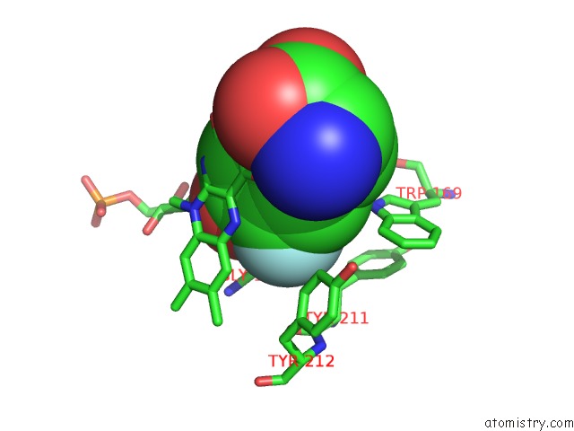
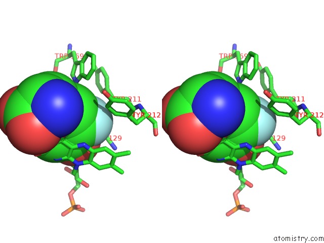
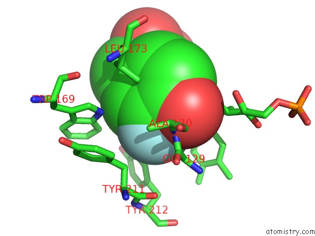
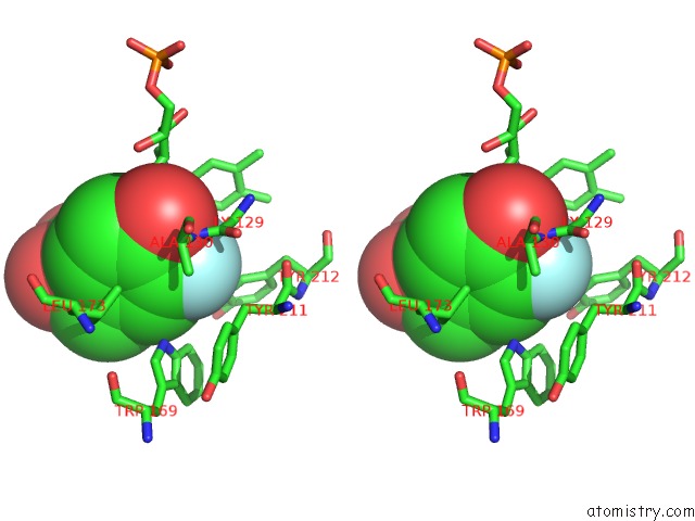
Fluorine binding site 1 out of 6 in 5yak
Go back to
Fluorine binding site 1 out
of 6 in the The Crystal Structure of Human Iyd THR239 Mutant with Ligand 3- Fluorotyrosine (F-Tyr)
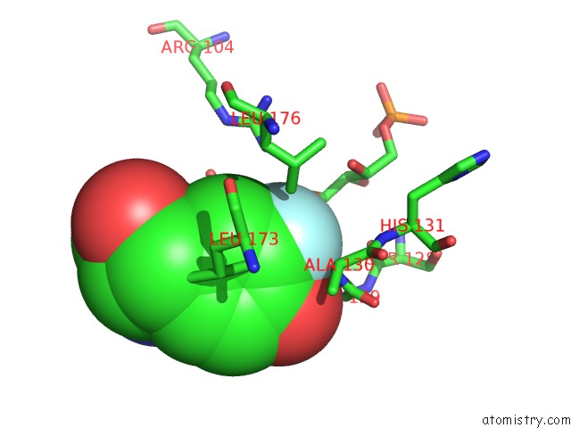
Mono view
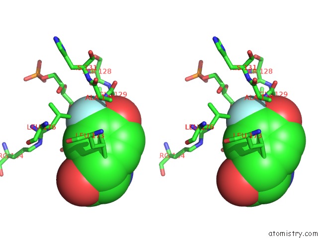
Stereo pair view
Mono view
Stereo pair view
A full contact list of Fluorine with other atoms in the F binding
site number 1 of The Crystal Structure of Human Iyd THR239 Mutant with Ligand 3- Fluorotyrosine (F-Tyr) within 5.0Å range:
|
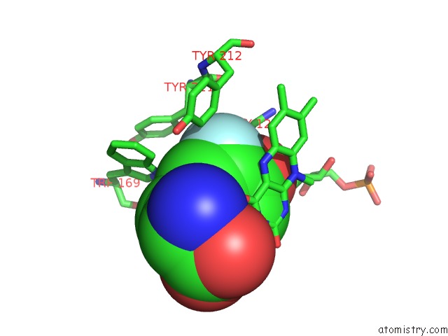
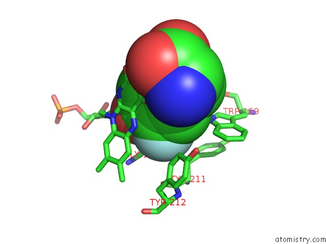
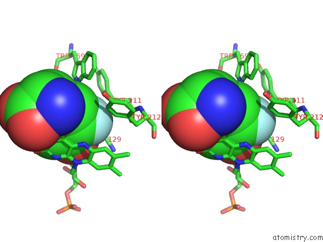
Fluorine binding site 2 out of 6 in 5yak
Go back to
Fluorine binding site 2 out
of 6 in the The Crystal Structure of Human Iyd THR239 Mutant with Ligand 3- Fluorotyrosine (F-Tyr)
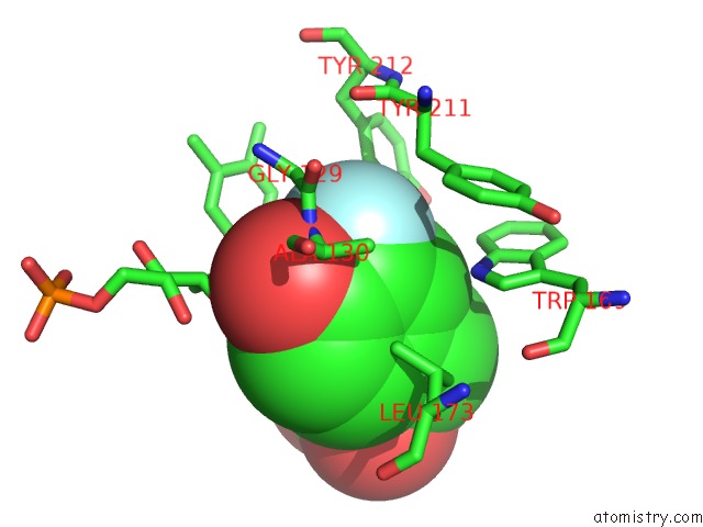
Mono view
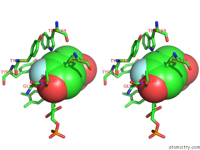
Stereo pair view
Mono view
Stereo pair view
A full contact list of Fluorine with other atoms in the F binding
site number 2 of The Crystal Structure of Human Iyd THR239 Mutant with Ligand 3- Fluorotyrosine (F-Tyr) within 5.0Å range:
|
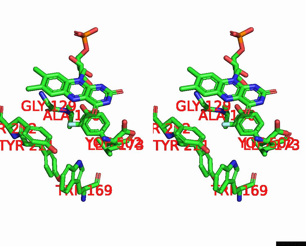
Fluorine binding site 3 out of 6 in 5yak
Go back to
Fluorine binding site 3 out
of 6 in the The Crystal Structure of Human Iyd THR239 Mutant with Ligand 3- Fluorotyrosine (F-Tyr)
Mono view
Stereo pair view
Mono view
Stereo pair view
A full contact list of Fluorine with other atoms in the F binding
site number 3 of The Crystal Structure of Human Iyd THR239 Mutant with Ligand 3- Fluorotyrosine (F-Tyr) within 5.0Å range:
|
Fluorine binding site 4 out of 6 in 5yak
Go back to
Fluorine binding site 4 out
of 6 in the The Crystal Structure of Human Iyd THR239 Mutant with Ligand 3- Fluorotyrosine (F-Tyr)
Mono view
Stereo pair view
Mono view
Stereo pair view
A full contact list of Fluorine with other atoms in the F binding
site number 4 of The Crystal Structure of Human Iyd THR239 Mutant with Ligand 3- Fluorotyrosine (F-Tyr) within 5.0Å range:
|
Fluorine binding site 5 out of 6 in 5yak
Go back to
Fluorine binding site 5 out
of 6 in the The Crystal Structure of Human Iyd THR239 Mutant with Ligand 3- Fluorotyrosine (F-Tyr)
Mono view
Stereo pair view
Mono view
Stereo pair view
A full contact list of Fluorine with other atoms in the F binding
site number 5 of The Crystal Structure of Human Iyd THR239 Mutant with Ligand 3- Fluorotyrosine (F-Tyr) within 5.0Å range:
|
Fluorine binding site 6 out of 6 in 5yak
Go back to
Fluorine binding site 6 out
of 6 in the The Crystal Structure of Human Iyd THR239 Mutant with Ligand 3- Fluorotyrosine (F-Tyr)
Mono view
Stereo pair view
Mono view
Stereo pair view
A full contact list of Fluorine with other atoms in the F binding
site number 6 of The Crystal Structure of Human Iyd THR239 Mutant with Ligand 3- Fluorotyrosine (F-Tyr) within 5.0Å range:
|
Reference:
J.Hu,
Q.Su,
J.L.Schlessman,
S.E.Rokita.
Redox Control of Iodotyrosine Deiodinase Protein Sci. V. 28 68 2019.
ISSN: ESSN 1469-896X
PubMed: 30052294
DOI: 10.1002/PRO.3479
Page generated: Thu Aug 1 17:10:12 2024
ISSN: ESSN 1469-896X
PubMed: 30052294
DOI: 10.1002/PRO.3479
Last articles
Zn in 9JYWZn in 9IR4
Zn in 9IR3
Zn in 9GMX
Zn in 9GMW
Zn in 9JEJ
Zn in 9ERF
Zn in 9ERE
Zn in 9EGV
Zn in 9EGW