Fluorine »
PDB 5y6d-5zab »
5ylu »
Fluorine in PDB 5ylu: Crystal Structure of the Gastric Proton Pump Complexed with Vonoprazan
Enzymatic activity of Crystal Structure of the Gastric Proton Pump Complexed with Vonoprazan
All present enzymatic activity of Crystal Structure of the Gastric Proton Pump Complexed with Vonoprazan:
3.6.3.10;
3.6.3.10;
Protein crystallography data
The structure of Crystal Structure of the Gastric Proton Pump Complexed with Vonoprazan, PDB code: 5ylu
was solved by
K.Abe,
K.Irie,
H.Nakanishi,
Y.Fujiyoshi,
with X-Ray Crystallography technique. A brief refinement statistics is given in the table below:
| Resolution Low / High (Å) | 48.18 / 2.80 |
| Space group | P 31 2 1 |
| Cell size a, b, c (Å), α, β, γ (°) | 104.820, 104.820, 367.080, 90.00, 90.00, 120.00 |
| R / Rfree (%) | 23.7 / 28.8 |
Other elements in 5ylu:
The structure of Crystal Structure of the Gastric Proton Pump Complexed with Vonoprazan also contains other interesting chemical elements:
| Magnesium | (Mg) | 1 atom |
Fluorine Binding Sites:
The binding sites of Fluorine atom in the Crystal Structure of the Gastric Proton Pump Complexed with Vonoprazan
(pdb code 5ylu). This binding sites where shown within
5.0 Angstroms radius around Fluorine atom.
In total 4 binding sites of Fluorine where determined in the Crystal Structure of the Gastric Proton Pump Complexed with Vonoprazan, PDB code: 5ylu:
Jump to Fluorine binding site number: 1; 2; 3; 4;
In total 4 binding sites of Fluorine where determined in the Crystal Structure of the Gastric Proton Pump Complexed with Vonoprazan, PDB code: 5ylu:
Jump to Fluorine binding site number: 1; 2; 3; 4;
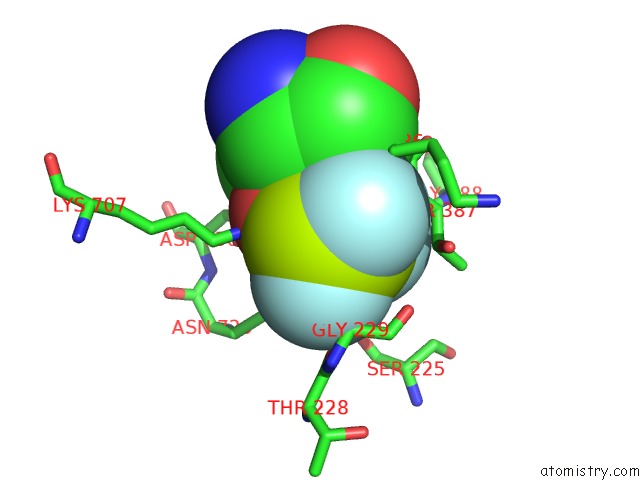
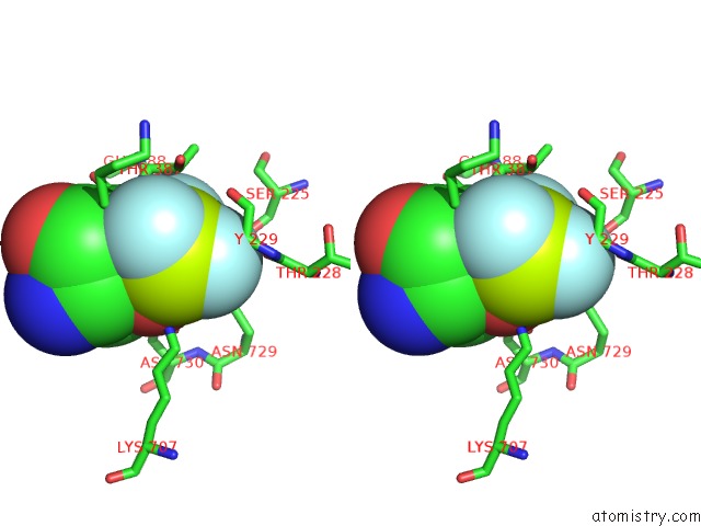
Fluorine binding site 1 out of 4 in 5ylu
Go back to
Fluorine binding site 1 out
of 4 in the Crystal Structure of the Gastric Proton Pump Complexed with Vonoprazan
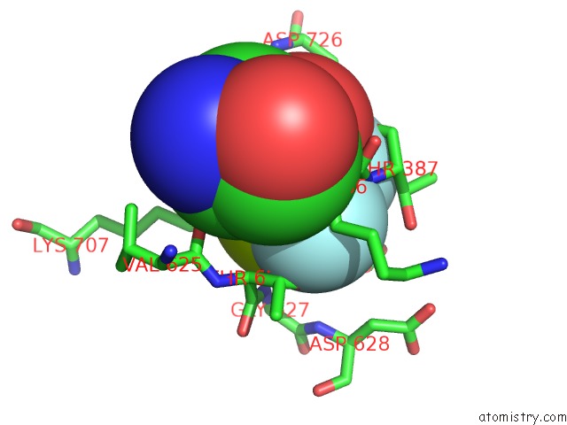
Mono view
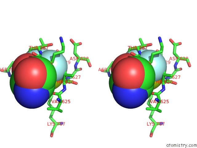
Stereo pair view
Mono view
Stereo pair view
A full contact list of Fluorine with other atoms in the F binding
site number 1 of Crystal Structure of the Gastric Proton Pump Complexed with Vonoprazan within 5.0Å range:
|
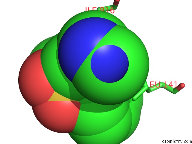
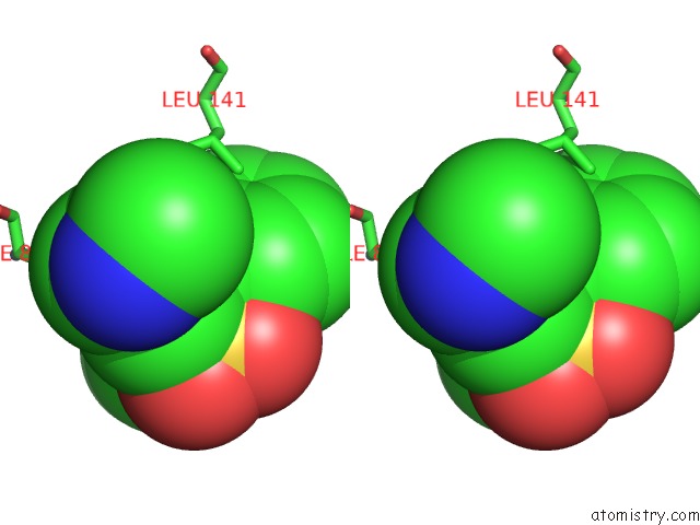
Fluorine binding site 2 out of 4 in 5ylu
Go back to
Fluorine binding site 2 out
of 4 in the Crystal Structure of the Gastric Proton Pump Complexed with Vonoprazan
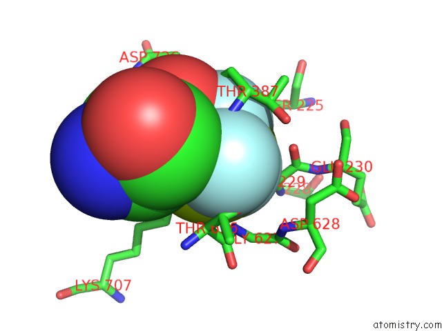
Mono view
Stereo pair view
Mono view
Stereo pair view
A full contact list of Fluorine with other atoms in the F binding
site number 2 of Crystal Structure of the Gastric Proton Pump Complexed with Vonoprazan within 5.0Å range:
|
Fluorine binding site 3 out of 4 in 5ylu
Go back to
Fluorine binding site 3 out
of 4 in the Crystal Structure of the Gastric Proton Pump Complexed with Vonoprazan
Mono view
Stereo pair view
Mono view
Stereo pair view
A full contact list of Fluorine with other atoms in the F binding
site number 3 of Crystal Structure of the Gastric Proton Pump Complexed with Vonoprazan within 5.0Å range:
|
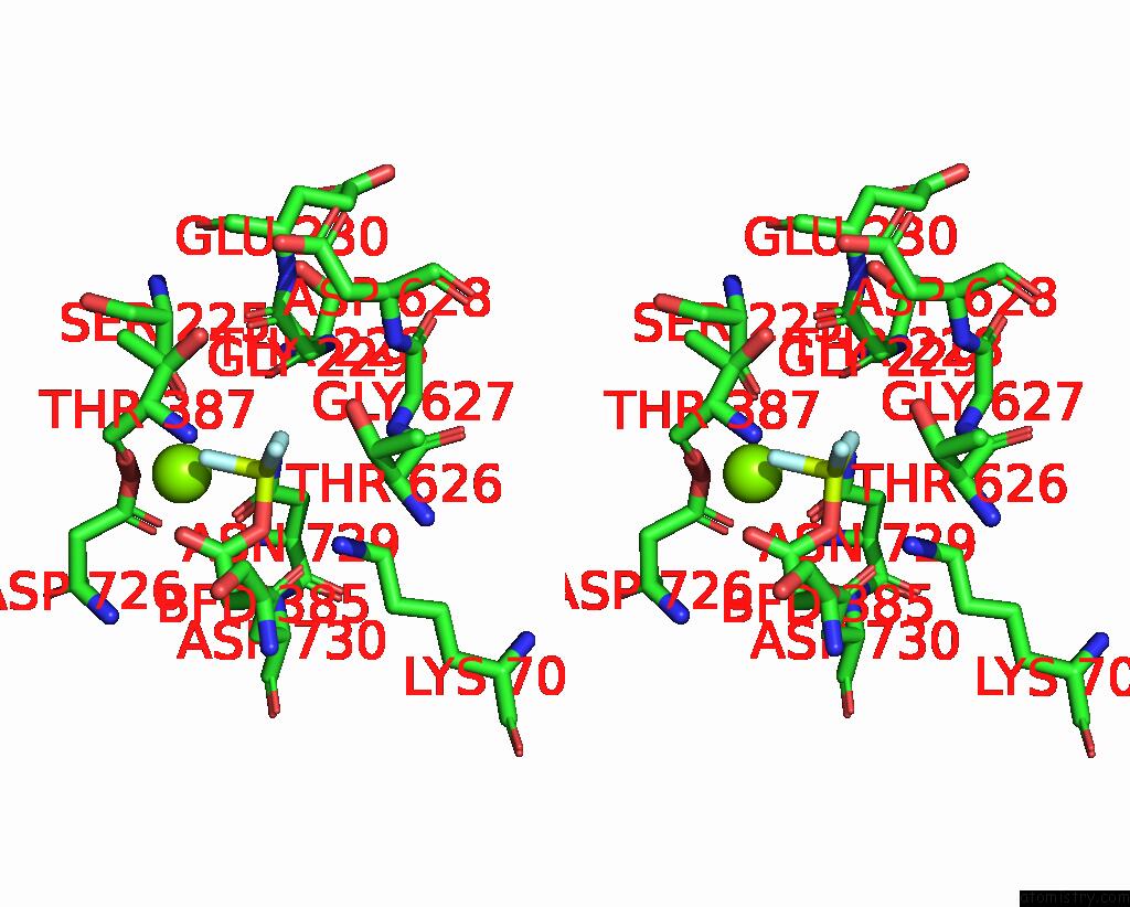
Fluorine binding site 4 out of 4 in 5ylu
Go back to
Fluorine binding site 4 out
of 4 in the Crystal Structure of the Gastric Proton Pump Complexed with Vonoprazan
Mono view
Stereo pair view
Mono view
Stereo pair view
A full contact list of Fluorine with other atoms in the F binding
site number 4 of Crystal Structure of the Gastric Proton Pump Complexed with Vonoprazan within 5.0Å range:
|
Reference:
K.Abe,
K.Irie,
H.Nakanishi,
H.Suzuki,
Y.Fujiyoshi.
Crystal Structures of the Gastric Proton Pump Nature V. 556 214 2018.
ISSN: ESSN 1476-4687
PubMed: 29618813
DOI: 10.1038/S41586-018-0003-8
Page generated: Thu Aug 1 17:20:17 2024
ISSN: ESSN 1476-4687
PubMed: 29618813
DOI: 10.1038/S41586-018-0003-8
Last articles
Zn in 9MJ5Zn in 9HNW
Zn in 9G0L
Zn in 9FNE
Zn in 9DZN
Zn in 9E0I
Zn in 9D32
Zn in 9DAK
Zn in 8ZXC
Zn in 8ZUF