Fluorine in PDB 6a36: Mandelate Oxidase Mutant-Y128F with the 3-Fluoropyruvic Acid Fmn Adduct
Enzymatic activity of Mandelate Oxidase Mutant-Y128F with the 3-Fluoropyruvic Acid Fmn Adduct
All present enzymatic activity of Mandelate Oxidase Mutant-Y128F with the 3-Fluoropyruvic Acid Fmn Adduct:
1.1.3.46;
Protein crystallography data
The structure of Mandelate Oxidase Mutant-Y128F with the 3-Fluoropyruvic Acid Fmn Adduct, PDB code: 6a36
was solved by
T.L.Li,
K.H.Lin,
with X-Ray Crystallography technique. A brief refinement statistics is given in the table below:
Resolution Low / High (Å)
|
30.00 /
1.44
|
Space group
|
I 4 2 2
|
Cell size a, b, c (Å), α, β, γ (°)
|
137.749,
137.749,
111.876,
90.00,
90.00,
90.00
|
R / Rfree (%)
|
17.6 /
19.2
|
Fluorine Binding Sites:
The binding sites of Fluorine atom in the Mandelate Oxidase Mutant-Y128F with the 3-Fluoropyruvic Acid Fmn Adduct
(pdb code 6a36). This binding sites where shown within
5.0 Angstroms radius around Fluorine atom.
In total 2 binding sites of Fluorine where determined in the
Mandelate Oxidase Mutant-Y128F with the 3-Fluoropyruvic Acid Fmn Adduct, PDB code: 6a36:
Jump to Fluorine binding site number:
1;
2;
Fluorine binding site 1 out
of 2 in 6a36
Go back to
Fluorine Binding Sites List in 6a36
Fluorine binding site 1 out
of 2 in the Mandelate Oxidase Mutant-Y128F with the 3-Fluoropyruvic Acid Fmn Adduct
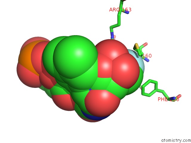
 Mono view
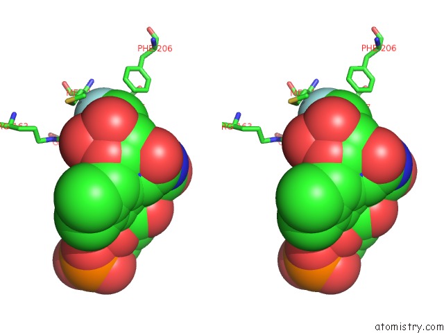
 Stereo pair view
|
A full contact list of Fluorine with other atoms in the F binding
site number 1 of Mandelate Oxidase Mutant-Y128F with the 3-Fluoropyruvic Acid Fmn Adduct within 5.0Å range:
probe
|
atom
|
residue
|
distance (Å)
|
B
|
Occ
|
A:F401
b:21.0
occ:0.50
|
FBK
|
A:9P9401
|
0.0
|
21.0
|
0.5
|
CBJ
|
A:9P9401
|
1.3
|
19.8
|
0.5
|
CBG
|
A:9P9401
|
2.5
|
18.2
|
0.5
|
OBO
|
A:9P9401
|
2.9
|
20.2
|
0.5
|
CBL
|
A:9P9401
|
3.0
|
18.7
|
0.5
|
CBH
|
A:9PF402
|
3.3
|
18.3
|
0.5
|
CG
|
A:MET160
|
3.3
|
27.7
|
1.0
|
CB
|
A:MET160
|
3.5
|
24.8
|
1.0
|
OBM
|
A:9P9401
|
3.5
|
19.6
|
0.5
|
OBN
|
A:9P9401
|
3.5
|
16.3
|
0.5
|
NH2
|
A:ARG163
|
3.6
|
18.9
|
1.0
|
FBI
|
A:9PF402
|
3.7
|
19.2
|
0.5
|
CE
|
A:MET160
|
3.7
|
33.0
|
1.0
|
CBF
|
A:9P9401
|
3.8
|
16.1
|
0.5
|
CA
|
A:MET160
|
3.8
|
21.4
|
1.0
|
CG1
|
A:VAL157
|
3.9
|
14.1
|
1.0
|
CZ
|
A:PHE206
|
4.1
|
42.8
|
1.0
|
OE1
|
A:GLN256
|
4.2
|
14.0
|
1.0
|
SD
|
A:MET160
|
4.2
|
32.5
|
1.0
|
CE2
|
A:PHE206
|
4.3
|
42.9
|
1.0
|
CBF
|
A:9PF402
|
4.4
|
18.2
|
0.5
|
CZ
|
A:ARG163
|
4.5
|
19.3
|
1.0
|
N
|
A:MET160
|
4.6
|
19.5
|
1.0
|
NH1
|
A:ARG163
|
4.7
|
20.7
|
1.0
|
CBH
|
A:9P9401
|
4.7
|
14.0
|
0.5
|
OBI
|
A:9P9401
|
4.7
|
15.6
|
0.5
|
CG2
|
A:VAL157
|
4.9
|
13.2
|
1.0
|
OBG
|
A:9PF402
|
4.9
|
19.2
|
0.5
|
CD
|
A:GLN256
|
4.9
|
12.3
|
1.0
|
CE1
|
A:PHE206
|
5.0
|
40.7
|
1.0
|
|
Fluorine binding site 2 out
of 2 in 6a36
Go back to
Fluorine Binding Sites List in 6a36
Fluorine binding site 2 out
of 2 in the Mandelate Oxidase Mutant-Y128F with the 3-Fluoropyruvic Acid Fmn Adduct
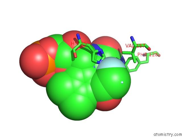
 Mono view
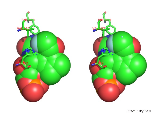
 Stereo pair view
|
A full contact list of Fluorine with other atoms in the F binding
site number 2 of Mandelate Oxidase Mutant-Y128F with the 3-Fluoropyruvic Acid Fmn Adduct within 5.0Å range:
probe
|
atom
|
residue
|
distance (Å)
|
B
|
Occ
|
A:F402
b:19.2
occ:0.50
|
FBI
|
A:9PF402
|
0.0
|
19.2
|
0.5
|
CBH
|
A:9PF402
|
1.1
|
18.3
|
0.5
|
CBF
|
A:9P9401
|
1.8
|
16.1
|
0.5
|
CBF
|
A:9PF402
|
2.2
|
18.2
|
0.5
|
CBG
|
A:9P9401
|
2.3
|
18.2
|
0.5
|
CBH
|
A:9P9401
|
2.5
|
14.0
|
0.5
|
CA
|
A:9PF402
|
2.5
|
15.1
|
0.5
|
CBJ
|
A:9P9401
|
2.6
|
19.8
|
0.5
|
C
|
A:9PF402
|
2.6
|
16.2
|
0.5
|
N
|
A:9PF402
|
2.7
|
16.4
|
0.5
|
OBN
|
A:9P9401
|
2.8
|
16.3
|
0.5
|
N
|
A:9P9401
|
2.9
|
10.7
|
0.5
|
C
|
A:9P9401
|
2.9
|
9.6
|
0.5
|
NE2
|
A:HIS252
|
2.9
|
12.4
|
1.0
|
O
|
A:9PF402
|
2.9
|
17.7
|
0.5
|
CBL
|
A:9P9401
|
2.9
|
18.7
|
0.5
|
CA
|
A:9P9401
|
2.9
|
9.8
|
0.5
|
CE1
|
A:HIS252
|
3.2
|
12.2
|
1.0
|
CAH
|
A:9P9401
|
3.3
|
8.9
|
0.5
|
NAS
|
A:9PF402
|
3.3
|
16.1
|
0.5
|
CAX
|
A:9PF402
|
3.3
|
14.7
|
0.5
|
O
|
A:9P9401
|
3.3
|
9.3
|
0.5
|
CZ
|
A:PHE128
|
3.4
|
14.5
|
1.0
|
OBG
|
A:9PF402
|
3.4
|
19.2
|
0.5
|
OBI
|
A:9P9401
|
3.5
|
15.6
|
0.5
|
NAM
|
A:9P9401
|
3.5
|
8.9
|
0.5
|
OBO
|
A:9P9401
|
3.6
|
20.2
|
0.5
|
FBK
|
A:9P9401
|
3.7
|
21.0
|
0.5
|
NAK
|
A:9P9401
|
3.7
|
8.7
|
0.5
|
CBA
|
A:9PF402
|
3.8
|
15.8
|
0.5
|
NAU
|
A:9PF402
|
3.8
|
15.1
|
0.5
|
CAL
|
A:9P9401
|
3.8
|
9.0
|
0.5
|
CAT
|
A:9PF402
|
3.8
|
15.8
|
0.5
|
CAE
|
A:9P9401
|
4.0
|
9.7
|
0.5
|
CE2
|
A:PHE128
|
4.0
|
14.6
|
1.0
|
NAG
|
A:9P9401
|
4.0
|
8.3
|
0.5
|
NAY
|
A:9PF402
|
4.1
|
14.1
|
0.5
|
OBM
|
A:9P9401
|
4.1
|
19.6
|
0.5
|
CD2
|
A:HIS252
|
4.2
|
11.9
|
1.0
|
CBB
|
A:9PF402
|
4.3
|
14.8
|
0.5
|
CAD
|
A:9P9401
|
4.4
|
8.9
|
0.5
|
CE1
|
A:PHE128
|
4.4
|
14.2
|
1.0
|
ND1
|
A:HIS252
|
4.5
|
11.6
|
1.0
|
CG2
|
A:VAL157
|
4.6
|
13.2
|
1.0
|
OAQ
|
A:9P9401
|
4.8
|
9.2
|
0.5
|
CAZ
|
A:9PF402
|
4.8
|
15.9
|
0.5
|
OAO
|
A:9PF402
|
4.8
|
16.8
|
0.5
|
CAF
|
A:9P9401
|
5.0
|
9.5
|
0.5
|
|
Reference:
T.L.Li,
K.H.Lin.
The Crystal Structure of Mandelate Oxidase Mutant-Y128F with the 3-Fluoropyruvic Acid Fmn Adduct To Be Published.
Page generated: Thu Aug 1 17:36:00 2024
|