Fluorine »
PDB 6b3e-6bkw »
6b9l »
Fluorine in PDB 6b9l: Crystal Structure of EPHA2 with Peptide 135E2
Enzymatic activity of Crystal Structure of EPHA2 with Peptide 135E2
All present enzymatic activity of Crystal Structure of EPHA2 with Peptide 135E2:
2.7.10.1;
2.7.10.1;
Protein crystallography data
The structure of Crystal Structure of EPHA2 with Peptide 135E2, PDB code: 6b9l
was solved by
J.Song,
X.Tan,
with X-Ray Crystallography technique. A brief refinement statistics is given in the table below:
| Resolution Low / High (Å) | 47.08 / 3.20 |
| Space group | P 21 21 21 |
| Cell size a, b, c (Å), α, β, γ (°) | 89.268, 94.153, 134.464, 90.00, 90.00, 90.00 |
| R / Rfree (%) | 23.5 / 27.4 |
Other elements in 6b9l:
The structure of Crystal Structure of EPHA2 with Peptide 135E2 also contains other interesting chemical elements:
| Chlorine | (Cl) | 4 atoms |
Fluorine Binding Sites:
The binding sites of Fluorine atom in the Crystal Structure of EPHA2 with Peptide 135E2
(pdb code 6b9l). This binding sites where shown within
5.0 Angstroms radius around Fluorine atom.
In total 4 binding sites of Fluorine where determined in the Crystal Structure of EPHA2 with Peptide 135E2, PDB code: 6b9l:
Jump to Fluorine binding site number: 1; 2; 3; 4;
In total 4 binding sites of Fluorine where determined in the Crystal Structure of EPHA2 with Peptide 135E2, PDB code: 6b9l:
Jump to Fluorine binding site number: 1; 2; 3; 4;
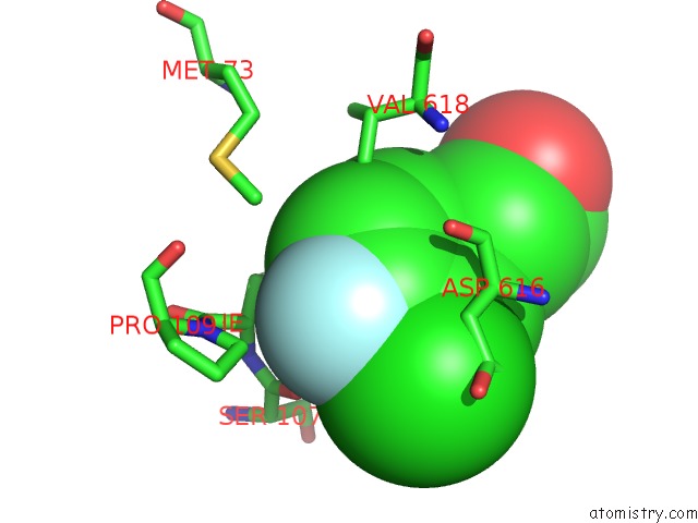
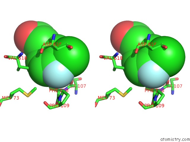
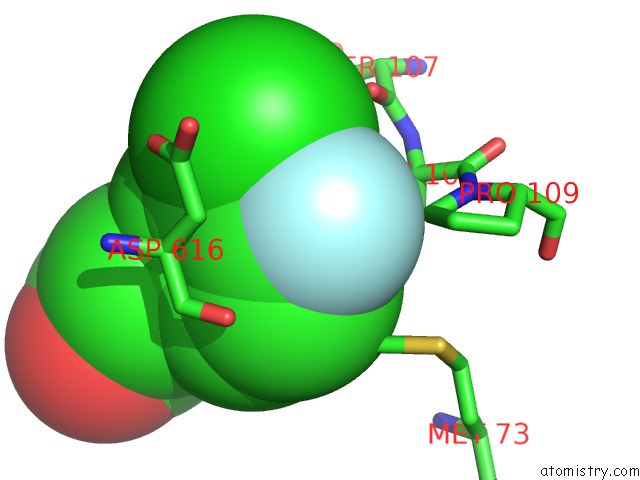
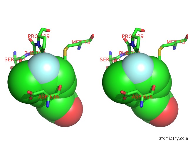
Fluorine binding site 1 out of 4 in 6b9l
Go back to
Fluorine binding site 1 out
of 4 in the Crystal Structure of EPHA2 with Peptide 135E2
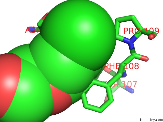
Mono view
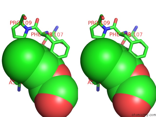
Stereo pair view
Mono view
Stereo pair view
A full contact list of Fluorine with other atoms in the F binding
site number 1 of Crystal Structure of EPHA2 with Peptide 135E2 within 5.0Å range:
|
Fluorine binding site 2 out of 4 in 6b9l
Go back to
Fluorine binding site 2 out
of 4 in the Crystal Structure of EPHA2 with Peptide 135E2
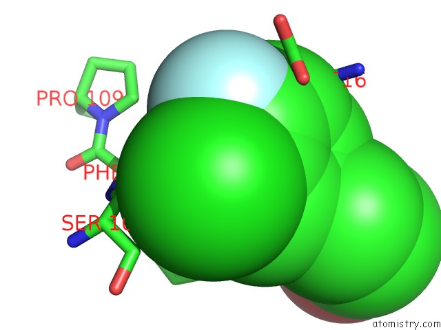
Mono view
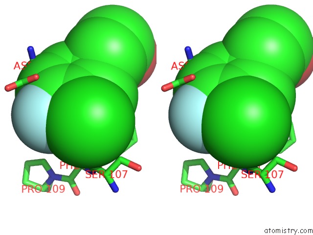
Stereo pair view
Mono view
Stereo pair view
A full contact list of Fluorine with other atoms in the F binding
site number 2 of Crystal Structure of EPHA2 with Peptide 135E2 within 5.0Å range:
|
Fluorine binding site 3 out of 4 in 6b9l
Go back to
Fluorine binding site 3 out
of 4 in the Crystal Structure of EPHA2 with Peptide 135E2
Mono view
Stereo pair view
Mono view
Stereo pair view
A full contact list of Fluorine with other atoms in the F binding
site number 3 of Crystal Structure of EPHA2 with Peptide 135E2 within 5.0Å range:
|
Fluorine binding site 4 out of 4 in 6b9l
Go back to
Fluorine binding site 4 out
of 4 in the Crystal Structure of EPHA2 with Peptide 135E2
Mono view
Stereo pair view
Mono view
Stereo pair view
A full contact list of Fluorine with other atoms in the F binding
site number 4 of Crystal Structure of EPHA2 with Peptide 135E2 within 5.0Å range:
|
Reference:
L.Gambini,
A.F.Salem,
P.Udompholkul,
X.F.Tan,
C.Baggio,
N.Shah,
A.Aronson,
J.Song,
M.Pellecchia.
Structure-Based Design of Novel EPHA2 Agonistic Agents with Nanomolar Affinity in Vitro and in Cell. Acs Chem. Biol. V. 13 2633 2018.
ISSN: ESSN 1554-8937
PubMed: 30110533
DOI: 10.1021/ACSCHEMBIO.8B00556
Page generated: Tue Jul 15 10:00:36 2025
ISSN: ESSN 1554-8937
PubMed: 30110533
DOI: 10.1021/ACSCHEMBIO.8B00556
Last articles
Fe in 2YXOFe in 2YRS
Fe in 2YXC
Fe in 2YNM
Fe in 2YVJ
Fe in 2YP1
Fe in 2YU2
Fe in 2YU1
Fe in 2YQB
Fe in 2YOO