Fluorine »
PDB 6bkw-6bz2 »
6bpv »
Fluorine in PDB 6bpv: Crystal Structure of Cysteine-Bound Ferrous Form of the Matured F2- TYR157 Human Cysteine Dioxygenase
Enzymatic activity of Crystal Structure of Cysteine-Bound Ferrous Form of the Matured F2- TYR157 Human Cysteine Dioxygenase
All present enzymatic activity of Crystal Structure of Cysteine-Bound Ferrous Form of the Matured F2- TYR157 Human Cysteine Dioxygenase:
1.13.11.20;
1.13.11.20;
Protein crystallography data
The structure of Crystal Structure of Cysteine-Bound Ferrous Form of the Matured F2- TYR157 Human Cysteine Dioxygenase, PDB code: 6bpv
was solved by
A.Liu,
J.Li,
I.Shin,
with X-Ray Crystallography technique. A brief refinement statistics is given in the table below:
| Resolution Low / High (Å) | 31.57 / 1.95 |
| Space group | P 65 |
| Cell size a, b, c (Å), α, β, γ (°) | 131.435, 131.435, 34.177, 90.00, 90.00, 120.00 |
| R / Rfree (%) | 16 / 17.9 |
Other elements in 6bpv:
The structure of Crystal Structure of Cysteine-Bound Ferrous Form of the Matured F2- TYR157 Human Cysteine Dioxygenase also contains other interesting chemical elements:
| Iron | (Fe) | 1 atom |
Fluorine Binding Sites:
The binding sites of Fluorine atom in the Crystal Structure of Cysteine-Bound Ferrous Form of the Matured F2- TYR157 Human Cysteine Dioxygenase
(pdb code 6bpv). This binding sites where shown within
5.0 Angstroms radius around Fluorine atom.
In total only one binding site of Fluorine was determined in the Crystal Structure of Cysteine-Bound Ferrous Form of the Matured F2- TYR157 Human Cysteine Dioxygenase, PDB code: 6bpv:
In total only one binding site of Fluorine was determined in the Crystal Structure of Cysteine-Bound Ferrous Form of the Matured F2- TYR157 Human Cysteine Dioxygenase, PDB code: 6bpv:
Fluorine binding site 1 out of 1 in 6bpv
Go back to
Fluorine binding site 1 out
of 1 in the Crystal Structure of Cysteine-Bound Ferrous Form of the Matured F2- TYR157 Human Cysteine Dioxygenase

Mono view
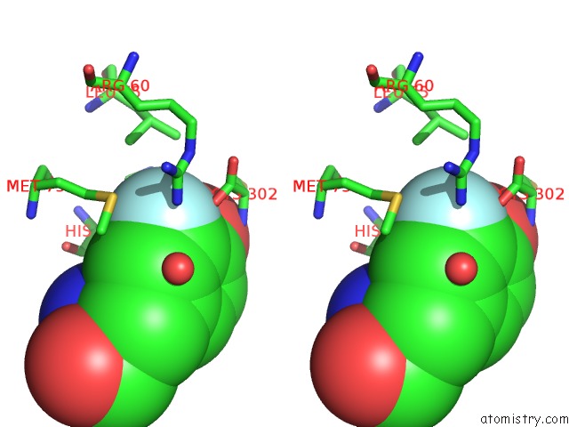
Stereo pair view
Mono view
Stereo pair view
A full contact list of Fluorine with other atoms in the F binding
site number 1 of Crystal Structure of Cysteine-Bound Ferrous Form of the Matured F2- TYR157 Human Cysteine Dioxygenase within 5.0Å range:
|
Reference:
J.Li,
W.P.Griffith,
I.Davis,
I.Shin,
J.Wang,
F.Li,
Y.Wang,
D.J.Wherritt,
A.Liu.
Cleavage of A Carbon-Fluorine Bond By An Engineered Cysteine Dioxygenase. Nat. Chem. Biol. V. 14 853 2018.
ISSN: ESSN 1552-4469
PubMed: 29942080
DOI: 10.1038/S41589-018-0085-5
Page generated: Thu Aug 1 18:14:55 2024
ISSN: ESSN 1552-4469
PubMed: 29942080
DOI: 10.1038/S41589-018-0085-5
Last articles
Zn in 9MJ5Zn in 9HNW
Zn in 9G0L
Zn in 9FNE
Zn in 9DZN
Zn in 9E0I
Zn in 9D32
Zn in 9DAK
Zn in 8ZXC
Zn in 8ZUF