Fluorine »
PDB 6eol-6fex »
6f6i »
Fluorine in PDB 6f6i: Crystal Structure of Ebolavirus Glycoprotein in Complex with Paroxetine
Protein crystallography data
The structure of Crystal Structure of Ebolavirus Glycoprotein in Complex with Paroxetine, PDB code: 6f6i
was solved by
J.Ren,
Y.Zhao,
E.E.Fry,
D.I.Stuart,
with X-Ray Crystallography technique. A brief refinement statistics is given in the table below:
| Resolution Low / High (Å) | 52.01 / 2.40 |
| Space group | H 3 2 |
| Cell size a, b, c (Å), α, β, γ (°) | 113.680, 113.680, 306.260, 90.00, 90.00, 120.00 |
| R / Rfree (%) | 19.2 / 21.2 |
Fluorine Binding Sites:
The binding sites of Fluorine atom in the Crystal Structure of Ebolavirus Glycoprotein in Complex with Paroxetine
(pdb code 6f6i). This binding sites where shown within
5.0 Angstroms radius around Fluorine atom.
In total only one binding site of Fluorine was determined in the Crystal Structure of Ebolavirus Glycoprotein in Complex with Paroxetine, PDB code: 6f6i:
In total only one binding site of Fluorine was determined in the Crystal Structure of Ebolavirus Glycoprotein in Complex with Paroxetine, PDB code: 6f6i:
Fluorine binding site 1 out of 1 in 6f6i
Go back to
Fluorine binding site 1 out
of 1 in the Crystal Structure of Ebolavirus Glycoprotein in Complex with Paroxetine
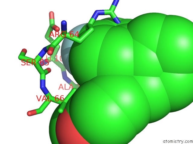
Mono view
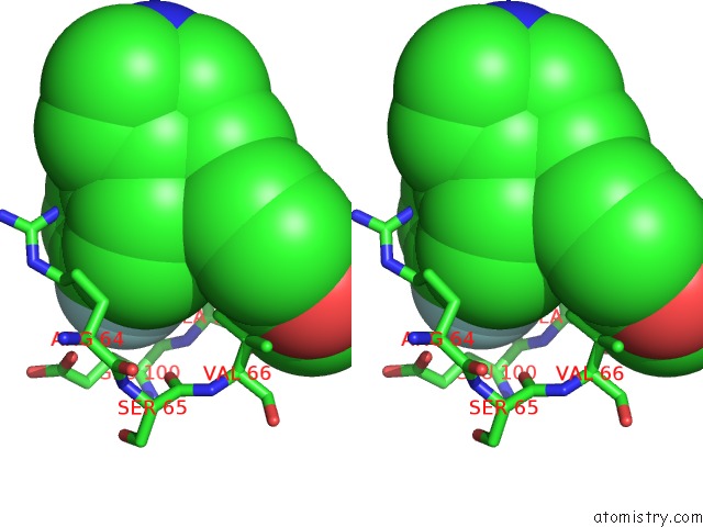
Stereo pair view
Mono view
Stereo pair view
A full contact list of Fluorine with other atoms in the F binding
site number 1 of Crystal Structure of Ebolavirus Glycoprotein in Complex with Paroxetine within 5.0Å range:
|
Reference:
J.Ren,
Y.Zhao,
E.E.Fry,
D.I.Stuart.
Target Identification and Mode of Action of Four Chemically Divergent Drugs Against Ebolavirus Infection. J. Med. Chem. V. 61 724 2018.
ISSN: ISSN 1520-4804
PubMed: 29272110
DOI: 10.1021/ACS.JMEDCHEM.7B01249
Page generated: Tue Jul 15 11:22:57 2025
ISSN: ISSN 1520-4804
PubMed: 29272110
DOI: 10.1021/ACS.JMEDCHEM.7B01249
Last articles
F in 6XOXF in 6XMU
F in 6XMT
F in 6XMS
F in 6XMK
F in 6XL4
F in 6XLO
F in 6XLS
F in 6XJU
F in 6XK9