Fluorine »
PDB 6gqm-6hax »
6h8r »
Fluorine in PDB 6h8r: Crystal Structure of the Human Protein Tyrosine Phosphatase PTPN5 (Step) in Complex with Compound 2
Enzymatic activity of Crystal Structure of the Human Protein Tyrosine Phosphatase PTPN5 (Step) in Complex with Compound 2
All present enzymatic activity of Crystal Structure of the Human Protein Tyrosine Phosphatase PTPN5 (Step) in Complex with Compound 2:
3.1.3.48;
3.1.3.48;
Protein crystallography data
The structure of Crystal Structure of the Human Protein Tyrosine Phosphatase PTPN5 (Step) in Complex with Compound 2, PDB code: 6h8r
was solved by
S.Hoerer,
D.Fiegen,
G.Schnapp,
with X-Ray Crystallography technique. A brief refinement statistics is given in the table below:
| Resolution Low / High (Å) | 39.69 / 1.66 |
| Space group | P 21 21 21 |
| Cell size a, b, c (Å), α, β, γ (°) | 53.095, 64.375, 100.804, 90.00, 90.00, 90.00 |
| R / Rfree (%) | 20 / 23.9 |
Fluorine Binding Sites:
The binding sites of Fluorine atom in the Crystal Structure of the Human Protein Tyrosine Phosphatase PTPN5 (Step) in Complex with Compound 2
(pdb code 6h8r). This binding sites where shown within
5.0 Angstroms radius around Fluorine atom.
In total 3 binding sites of Fluorine where determined in the Crystal Structure of the Human Protein Tyrosine Phosphatase PTPN5 (Step) in Complex with Compound 2, PDB code: 6h8r:
Jump to Fluorine binding site number: 1; 2; 3;
In total 3 binding sites of Fluorine where determined in the Crystal Structure of the Human Protein Tyrosine Phosphatase PTPN5 (Step) in Complex with Compound 2, PDB code: 6h8r:
Jump to Fluorine binding site number: 1; 2; 3;
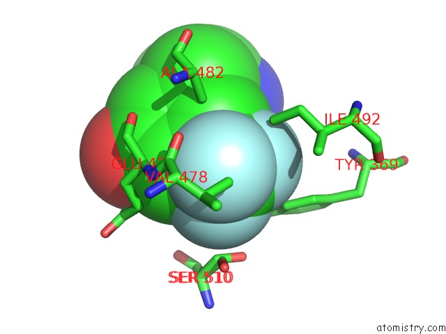
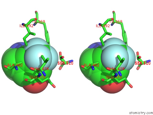
Fluorine binding site 1 out of 3 in 6h8r
Go back to
Fluorine binding site 1 out
of 3 in the Crystal Structure of the Human Protein Tyrosine Phosphatase PTPN5 (Step) in Complex with Compound 2
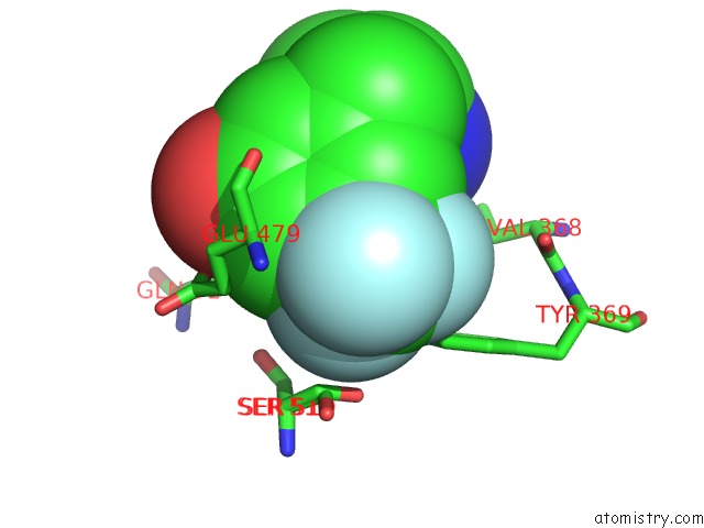
Mono view
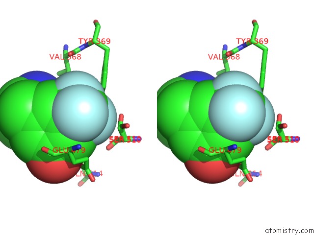
Stereo pair view
Mono view
Stereo pair view
A full contact list of Fluorine with other atoms in the F binding
site number 1 of Crystal Structure of the Human Protein Tyrosine Phosphatase PTPN5 (Step) in Complex with Compound 2 within 5.0Å range:
|
Fluorine binding site 2 out of 3 in 6h8r
Go back to
Fluorine binding site 2 out
of 3 in the Crystal Structure of the Human Protein Tyrosine Phosphatase PTPN5 (Step) in Complex with Compound 2
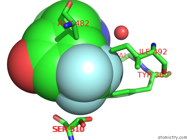
Mono view
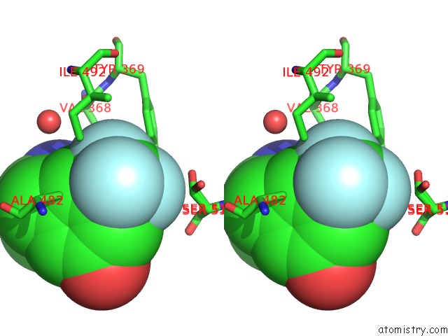
Stereo pair view
Mono view
Stereo pair view
A full contact list of Fluorine with other atoms in the F binding
site number 2 of Crystal Structure of the Human Protein Tyrosine Phosphatase PTPN5 (Step) in Complex with Compound 2 within 5.0Å range:
|
Fluorine binding site 3 out of 3 in 6h8r
Go back to
Fluorine binding site 3 out
of 3 in the Crystal Structure of the Human Protein Tyrosine Phosphatase PTPN5 (Step) in Complex with Compound 2
Mono view
Stereo pair view
Mono view
Stereo pair view
A full contact list of Fluorine with other atoms in the F binding
site number 3 of Crystal Structure of the Human Protein Tyrosine Phosphatase PTPN5 (Step) in Complex with Compound 2 within 5.0Å range:
|
Reference:
C.S.Tautermann,
F.Binder,
F.H.Buttner,
C.Eickmeier,
D.Fiegen,
U.Gross,
M.A.Grundl,
R.Heilker,
S.Hobson,
S.Hoerer,
A.Luippold,
V.Mack,
F.Montel,
S.Peters,
S.Bhattacharya,
N.Vaidehi,
G.Schnapp,
S.Thamm,
M.Zeeb.
Allosteric Activation of Striatal-Enriched Protein Tyrosine Phosphatase (Step, PTPN5) By A Fragment-Like Molecule. J. Med. Chem. V. 62 306 2019.
ISSN: ISSN 1520-4804
PubMed: 30207464
DOI: 10.1021/ACS.JMEDCHEM.8B00857
Page generated: Thu Aug 1 20:54:10 2024
ISSN: ISSN 1520-4804
PubMed: 30207464
DOI: 10.1021/ACS.JMEDCHEM.8B00857
Last articles
Zn in 9J0NZn in 9J0O
Zn in 9J0P
Zn in 9FJX
Zn in 9EKB
Zn in 9C0F
Zn in 9CAH
Zn in 9CH0
Zn in 9CH3
Zn in 9CH1