Fluorine »
PDB 6gqy-6hay »
6ha9 »
Fluorine in PDB 6ha9: Structure of An Endo-Xyloglucanase From Cellvibrio Japonicus Complexed with Xxxg(2F)-Beta-Dnp
Enzymatic activity of Structure of An Endo-Xyloglucanase From Cellvibrio Japonicus Complexed with Xxxg(2F)-Beta-Dnp
All present enzymatic activity of Structure of An Endo-Xyloglucanase From Cellvibrio Japonicus Complexed with Xxxg(2F)-Beta-Dnp:
3.2.1.151;
3.2.1.151;
Protein crystallography data
The structure of Structure of An Endo-Xyloglucanase From Cellvibrio Japonicus Complexed with Xxxg(2F)-Beta-Dnp, PDB code: 6ha9
was solved by
W.A.Offen,
G.J.Davies,
with X-Ray Crystallography technique. A brief refinement statistics is given in the table below:
| Resolution Low / High (Å) | 79.19 / 2.00 |
| Space group | P 21 21 21 |
| Cell size a, b, c (Å), α, β, γ (°) | 56.476, 97.809, 158.121, 90.00, 90.00, 90.00 |
| R / Rfree (%) | 21.9 / 28.7 |
Fluorine Binding Sites:
The binding sites of Fluorine atom in the Structure of An Endo-Xyloglucanase From Cellvibrio Japonicus Complexed with Xxxg(2F)-Beta-Dnp
(pdb code 6ha9). This binding sites where shown within
5.0 Angstroms radius around Fluorine atom.
In total 2 binding sites of Fluorine where determined in the Structure of An Endo-Xyloglucanase From Cellvibrio Japonicus Complexed with Xxxg(2F)-Beta-Dnp, PDB code: 6ha9:
Jump to Fluorine binding site number: 1; 2;
In total 2 binding sites of Fluorine where determined in the Structure of An Endo-Xyloglucanase From Cellvibrio Japonicus Complexed with Xxxg(2F)-Beta-Dnp, PDB code: 6ha9:
Jump to Fluorine binding site number: 1; 2;
Fluorine binding site 1 out of 2 in 6ha9
Go back to
Fluorine binding site 1 out
of 2 in the Structure of An Endo-Xyloglucanase From Cellvibrio Japonicus Complexed with Xxxg(2F)-Beta-Dnp
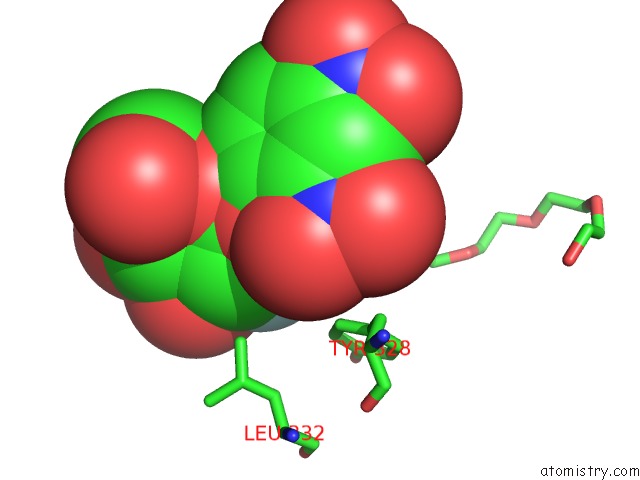
Mono view
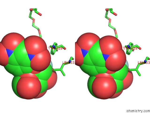
Stereo pair view
Mono view
Stereo pair view
A full contact list of Fluorine with other atoms in the F binding
site number 1 of Structure of An Endo-Xyloglucanase From Cellvibrio Japonicus Complexed with Xxxg(2F)-Beta-Dnp within 5.0Å range:
|
Fluorine binding site 2 out of 2 in 6ha9
Go back to
Fluorine binding site 2 out
of 2 in the Structure of An Endo-Xyloglucanase From Cellvibrio Japonicus Complexed with Xxxg(2F)-Beta-Dnp
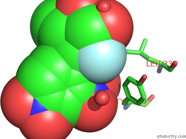
Mono view
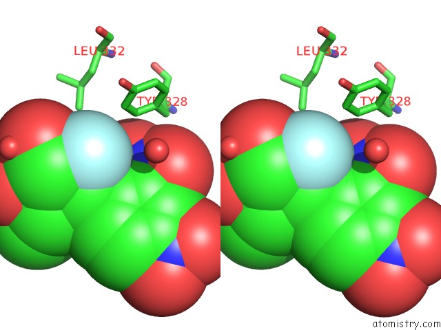
Stereo pair view
Mono view
Stereo pair view
A full contact list of Fluorine with other atoms in the F binding
site number 2 of Structure of An Endo-Xyloglucanase From Cellvibrio Japonicus Complexed with Xxxg(2F)-Beta-Dnp within 5.0Å range:
|
Reference:
N.Jain,
M.A.Attia,
W.A.Offen,
G.J.Davies,
H.Brumer.
Synthesis and Application of A Highly Branched, Mechanism-Based 2-Deoxy-2-Fluoro-Oligosaccharide Inhibitor of Endo-Xyloglucanases. Org. Biomol. Chem. V. 16 8732 2018.
ISSN: ESSN 1477-0539
PubMed: 30387796
DOI: 10.1039/C8OB02250J
Page generated: Tue Jul 15 12:09:29 2025
ISSN: ESSN 1477-0539
PubMed: 30387796
DOI: 10.1039/C8OB02250J
Last articles
Fe in 2YXOFe in 2YRS
Fe in 2YXC
Fe in 2YNM
Fe in 2YVJ
Fe in 2YP1
Fe in 2YU2
Fe in 2YU1
Fe in 2YQB
Fe in 2YOO