Fluorine »
PDB 6hay-6hmo »
6hkz »
Fluorine in PDB 6hkz: X-Ray Structure of Human Glutamate Carboxypeptidase II (Gcpii) in Complex with A Inhibitor Rna 2-49-1
Enzymatic activity of X-Ray Structure of Human Glutamate Carboxypeptidase II (Gcpii) in Complex with A Inhibitor Rna 2-49-1
All present enzymatic activity of X-Ray Structure of Human Glutamate Carboxypeptidase II (Gcpii) in Complex with A Inhibitor Rna 2-49-1:
3.4.17.21;
3.4.17.21;
Protein crystallography data
The structure of X-Ray Structure of Human Glutamate Carboxypeptidase II (Gcpii) in Complex with A Inhibitor Rna 2-49-1, PDB code: 6hkz
was solved by
L.Motlova,
Z.Novakova,
C.Barinka,
with X-Ray Crystallography technique. A brief refinement statistics is given in the table below:
| Resolution Low / High (Å) | 47.59 / 2.09 |
| Space group | I 2 2 2 |
| Cell size a, b, c (Å), α, β, γ (°) | 102.956, 132.259, 161.589, 90.00, 90.00, 90.00 |
| R / Rfree (%) | 17 / 20.3 |
Other elements in 6hkz:
The structure of X-Ray Structure of Human Glutamate Carboxypeptidase II (Gcpii) in Complex with A Inhibitor Rna 2-49-1 also contains other interesting chemical elements:
| Zinc | (Zn) | 2 atoms |
| Calcium | (Ca) | 1 atom |
| Chlorine | (Cl) | 1 atom |
Fluorine Binding Sites:
The binding sites of Fluorine atom in the X-Ray Structure of Human Glutamate Carboxypeptidase II (Gcpii) in Complex with A Inhibitor Rna 2-49-1
(pdb code 6hkz). This binding sites where shown within
5.0 Angstroms radius around Fluorine atom.
In total only one binding site of Fluorine was determined in the X-Ray Structure of Human Glutamate Carboxypeptidase II (Gcpii) in Complex with A Inhibitor Rna 2-49-1, PDB code: 6hkz:
In total only one binding site of Fluorine was determined in the X-Ray Structure of Human Glutamate Carboxypeptidase II (Gcpii) in Complex with A Inhibitor Rna 2-49-1, PDB code: 6hkz:
Fluorine binding site 1 out of 1 in 6hkz
Go back to
Fluorine binding site 1 out
of 1 in the X-Ray Structure of Human Glutamate Carboxypeptidase II (Gcpii) in Complex with A Inhibitor Rna 2-49-1
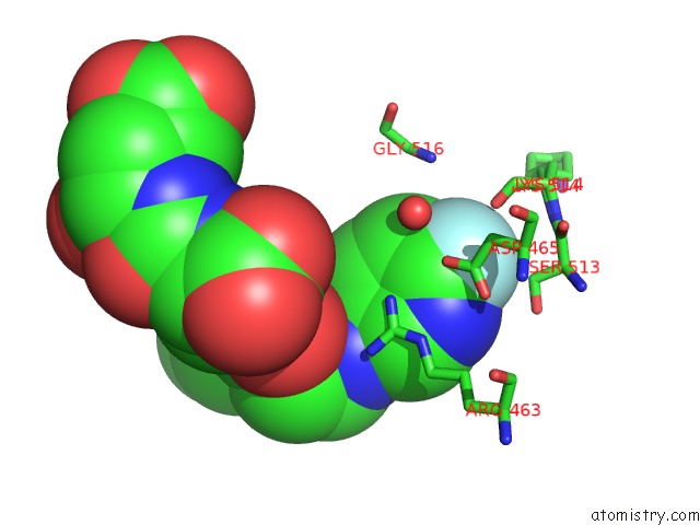
Mono view
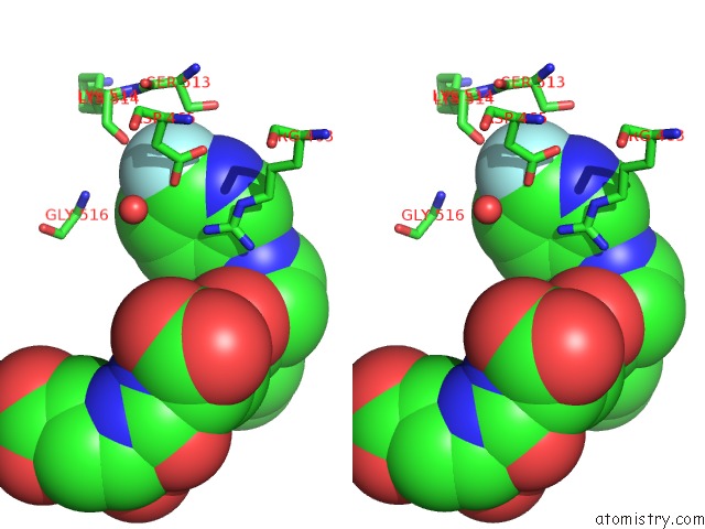
Stereo pair view
Mono view
Stereo pair view
A full contact list of Fluorine with other atoms in the F binding
site number 1 of X-Ray Structure of Human Glutamate Carboxypeptidase II (Gcpii) in Complex with A Inhibitor Rna 2-49-1 within 5.0Å range:
|
Reference:
R.Nakajima,
Z.Novakova,
W.Tueckmantel,
L.Motlova,
C.Barinka,
A.P.Kozikowski.
2-Aminoadipic Acid-C(O)-Glutamate Based Prostate-Specific Membrane Antigen Ligands For Potential Use As Theranostics. Acs Med Chem Lett V. 9 1099 2018.
ISSN: ISSN 1948-5875
PubMed: 30429952
DOI: 10.1021/ACSMEDCHEMLETT.8B00318
Page generated: Thu Aug 1 21:11:48 2024
ISSN: ISSN 1948-5875
PubMed: 30429952
DOI: 10.1021/ACSMEDCHEMLETT.8B00318
Last articles
Zn in 9MJ5Zn in 9HNW
Zn in 9G0L
Zn in 9FNE
Zn in 9DZN
Zn in 9E0I
Zn in 9D32
Zn in 9DAK
Zn in 8ZXC
Zn in 8ZUF