Fluorine »
PDB 6jsg-6kk1 »
6jtg »
Fluorine in PDB 6jtg: Structural Insights Into G Domain Dimerization and Pathogenic Mutations of OPA1
Enzymatic activity of Structural Insights Into G Domain Dimerization and Pathogenic Mutations of OPA1
All present enzymatic activity of Structural Insights Into G Domain Dimerization and Pathogenic Mutations of OPA1:
3.6.5.5;
3.6.5.5;
Protein crystallography data
The structure of Structural Insights Into G Domain Dimerization and Pathogenic Mutations of OPA1, PDB code: 6jtg
was solved by
L.Yan,
J.Hu,
with X-Ray Crystallography technique. A brief refinement statistics is given in the table below:
| Resolution Low / High (Å) | 46.35 / 2.40 |
| Space group | P 43 21 2 |
| Cell size a, b, c (Å), α, β, γ (°) | 77.890, 77.890, 171.547, 90.00, 90.00, 90.00 |
| R / Rfree (%) | 19.9 / 24.4 |
Other elements in 6jtg:
The structure of Structural Insights Into G Domain Dimerization and Pathogenic Mutations of OPA1 also contains other interesting chemical elements:
| Magnesium | (Mg) | 1 atom |
| Potassium | (K) | 1 atom |
Fluorine Binding Sites:
The binding sites of Fluorine atom in the Structural Insights Into G Domain Dimerization and Pathogenic Mutations of OPA1
(pdb code 6jtg). This binding sites where shown within
5.0 Angstroms radius around Fluorine atom.
In total 3 binding sites of Fluorine where determined in the Structural Insights Into G Domain Dimerization and Pathogenic Mutations of OPA1, PDB code: 6jtg:
Jump to Fluorine binding site number: 1; 2; 3;
In total 3 binding sites of Fluorine where determined in the Structural Insights Into G Domain Dimerization and Pathogenic Mutations of OPA1, PDB code: 6jtg:
Jump to Fluorine binding site number: 1; 2; 3;
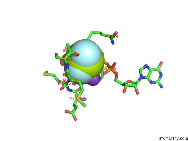
Fluorine binding site 1 out of 3 in 6jtg
Go back to
Fluorine binding site 1 out
of 3 in the Structural Insights Into G Domain Dimerization and Pathogenic Mutations of OPA1
Mono view

Stereo pair view
Mono view
Stereo pair view
A full contact list of Fluorine with other atoms in the F binding
site number 1 of Structural Insights Into G Domain Dimerization and Pathogenic Mutations of OPA1 within 5.0Å range:
|
Fluorine binding site 2 out of 3 in 6jtg
Go back to
Fluorine binding site 2 out
of 3 in the Structural Insights Into G Domain Dimerization and Pathogenic Mutations of OPA1

Mono view

Stereo pair view
Mono view
Stereo pair view
A full contact list of Fluorine with other atoms in the F binding
site number 2 of Structural Insights Into G Domain Dimerization and Pathogenic Mutations of OPA1 within 5.0Å range:
|
Fluorine binding site 3 out of 3 in 6jtg
Go back to
Fluorine binding site 3 out
of 3 in the Structural Insights Into G Domain Dimerization and Pathogenic Mutations of OPA1

Mono view

Stereo pair view
Mono view
Stereo pair view
A full contact list of Fluorine with other atoms in the F binding
site number 3 of Structural Insights Into G Domain Dimerization and Pathogenic Mutations of OPA1 within 5.0Å range:
|
Reference:
L.Yan,
J.Hu.
Structural Insights Into G Domain Dimerization and Pathogenic Mutations of OPA1 To Be Published.
Page generated: Tue Jul 15 12:42:55 2025
Last articles
F in 7FQLF in 7FTO
F in 7FT0
F in 7FTI
F in 7FQK
F in 7FSV
F in 7FSZ
F in 7FQZ
F in 7FQJ
F in 7FQI