Fluorine »
PDB 6mii-6n4b »
6mlw »
Fluorine in PDB 6mlw: Crystal Structure of X. Citri Phosphoglucomutase in Complex with 2- Fluoro Mannosyl-1-Methyl-Phosphonic Acid
Protein crystallography data
The structure of Crystal Structure of X. Citri Phosphoglucomutase in Complex with 2- Fluoro Mannosyl-1-Methyl-Phosphonic Acid, PDB code: 6mlw
was solved by
L.Beamer,
K.Stiers,
with X-Ray Crystallography technique. A brief refinement statistics is given in the table below:
| Resolution Low / High (Å) | 42.44 / 1.90 |
| Space group | P 21 21 21 |
| Cell size a, b, c (Å), α, β, γ (°) | 43.781, 54.587, 172.909, 90.00, 90.00, 90.00 |
| R / Rfree (%) | 18.9 / 24.9 |
Other elements in 6mlw:
The structure of Crystal Structure of X. Citri Phosphoglucomutase in Complex with 2- Fluoro Mannosyl-1-Methyl-Phosphonic Acid also contains other interesting chemical elements:
| Magnesium | (Mg) | 1 atom |
Fluorine Binding Sites:
The binding sites of Fluorine atom in the Crystal Structure of X. Citri Phosphoglucomutase in Complex with 2- Fluoro Mannosyl-1-Methyl-Phosphonic Acid
(pdb code 6mlw). This binding sites where shown within
5.0 Angstroms radius around Fluorine atom.
In total only one binding site of Fluorine was determined in the Crystal Structure of X. Citri Phosphoglucomutase in Complex with 2- Fluoro Mannosyl-1-Methyl-Phosphonic Acid, PDB code: 6mlw:
In total only one binding site of Fluorine was determined in the Crystal Structure of X. Citri Phosphoglucomutase in Complex with 2- Fluoro Mannosyl-1-Methyl-Phosphonic Acid, PDB code: 6mlw:
Fluorine binding site 1 out of 1 in 6mlw
Go back to
Fluorine binding site 1 out
of 1 in the Crystal Structure of X. Citri Phosphoglucomutase in Complex with 2- Fluoro Mannosyl-1-Methyl-Phosphonic Acid
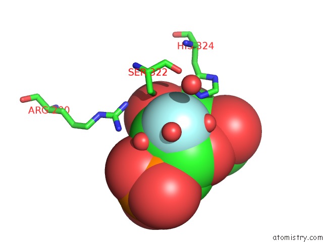
Mono view
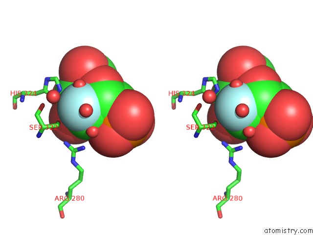
Stereo pair view
Mono view
Stereo pair view
A full contact list of Fluorine with other atoms in the F binding
site number 1 of Crystal Structure of X. Citri Phosphoglucomutase in Complex with 2- Fluoro Mannosyl-1-Methyl-Phosphonic Acid within 5.0Å range:
|
Reference:
J.S.Zhu,
K.M.Stiers,
E.Soleimani,
B.R.Groves,
L.J.Beamer,
D.L.Jakeman.
Inhibitory Evaluation of Alpha Pmm/Pgm Frompseudomonas Aeruginosa: Chemical Synthesis, Enzyme Kinetics, and Protein Crystallographic Study. J.Org.Chem. V. 84 9627 2019.
ISSN: ISSN 0022-3263
PubMed: 31264865
DOI: 10.1021/ACS.JOC.9B01305
Page generated: Thu Aug 1 22:19:02 2024
ISSN: ISSN 0022-3263
PubMed: 31264865
DOI: 10.1021/ACS.JOC.9B01305
Last articles
Zn in 9MJ5Zn in 9HNW
Zn in 9G0L
Zn in 9FNE
Zn in 9DZN
Zn in 9E0I
Zn in 9D32
Zn in 9DAK
Zn in 8ZXC
Zn in 8ZUF