Fluorine »
PDB 6mii-6n4b »
6mob »
Fluorine in PDB 6mob: Crystal Structure of KIT1 in Complex with DP2976 Via Co- Crystallization
Enzymatic activity of Crystal Structure of KIT1 in Complex with DP2976 Via Co- Crystallization
All present enzymatic activity of Crystal Structure of KIT1 in Complex with DP2976 Via Co- Crystallization:
2.7.10.1;
2.7.10.1;
Protein crystallography data
The structure of Crystal Structure of KIT1 in Complex with DP2976 Via Co- Crystallization, PDB code: 6mob
was solved by
T.E.Edwards,
J.Abendroth,
K.Safford,
L.Chun,
with X-Ray Crystallography technique. A brief refinement statistics is given in the table below:
| Resolution Low / High (Å) | 50.00 / 1.80 |
| Space group | P 43 21 2 |
| Cell size a, b, c (Å), α, β, γ (°) | 80.699, 80.699, 145.020, 90.00, 90.00, 90.00 |
| R / Rfree (%) | 18.1 / 21 |
Other elements in 6mob:
The structure of Crystal Structure of KIT1 in Complex with DP2976 Via Co- Crystallization also contains other interesting chemical elements:
| Chlorine | (Cl) | 1 atom |
Fluorine Binding Sites:
The binding sites of Fluorine atom in the Crystal Structure of KIT1 in Complex with DP2976 Via Co- Crystallization
(pdb code 6mob). This binding sites where shown within
5.0 Angstroms radius around Fluorine atom.
In total only one binding site of Fluorine was determined in the Crystal Structure of KIT1 in Complex with DP2976 Via Co- Crystallization, PDB code: 6mob:
In total only one binding site of Fluorine was determined in the Crystal Structure of KIT1 in Complex with DP2976 Via Co- Crystallization, PDB code: 6mob:
Fluorine binding site 1 out of 1 in 6mob
Go back to
Fluorine binding site 1 out
of 1 in the Crystal Structure of KIT1 in Complex with DP2976 Via Co- Crystallization
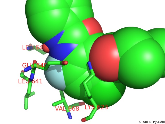
Mono view
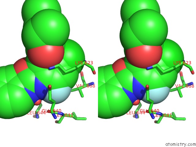
Stereo pair view
Mono view
Stereo pair view
A full contact list of Fluorine with other atoms in the F binding
site number 1 of Crystal Structure of KIT1 in Complex with DP2976 Via Co- Crystallization within 5.0Å range:
|
Reference:
B.D.Smith,
M.D.Kaufman,
W.P.Lu,
A.Gupta,
C.B.Leary,
S.C.Wise,
T.J.Rutkoski,
Y.M.Ahn,
G.Al-Ani,
S.L.Bulfer,
T.M.Caldwell,
L.Chun,
C.L.Ensinger,
M.M.Hood,
A.Mckinley,
W.C.Patt,
R.Ruiz-Soto,
Y.Su,
H.Telikepalli,
A.Town,
B.A.Turner,
L.Vogeti,
S.Vogeti,
K.Yates,
F.Janku,
A.R.Abdul Razak,
O.Rosen,
M.C.Heinrich,
D.L.Flynn.
Ripretinib (Dcc-2618) Is A Switch Control Kinase Inhibitor of A Broad Spectrum of Oncogenic and Drug-Resistant Kit and Pdgfra Variants. Cancer Cell V. 35 738 2019.
ISSN: ISSN 1535-6108
PubMed: 31085175
DOI: 10.1016/J.CCELL.2019.04.006
Page generated: Thu Aug 1 22:19:02 2024
ISSN: ISSN 1535-6108
PubMed: 31085175
DOI: 10.1016/J.CCELL.2019.04.006
Last articles
Zn in 9J0NZn in 9J0O
Zn in 9J0P
Zn in 9FJX
Zn in 9EKB
Zn in 9C0F
Zn in 9CAH
Zn in 9CH0
Zn in 9CH3
Zn in 9CH1