Fluorine »
PDB 6mjq-6n4e »
6n3z »
Fluorine in PDB 6n3z: Crystal Structure of An Epoxide Hydrolase From Trichoderma Reesei in Complex with Inhibitor 4
Protein crystallography data
The structure of Crystal Structure of An Epoxide Hydrolase From Trichoderma Reesei in Complex with Inhibitor 4, PDB code: 6n3z
was solved by
G.S.De Oliveira,
P.P.Adriani,
J.A.Ribeiro,
C.Morisseau,
B.D.Hammock,
M.V.Dias,
F.S.Chambergo,
with X-Ray Crystallography technique. A brief refinement statistics is given in the table below:
| Resolution Low / High (Å) | 38.06 / 2.24 |
| Space group | P 2 21 21 |
| Cell size a, b, c (Å), α, β, γ (°) | 50.527, 77.635, 86.827, 90.00, 90.00, 90.00 |
| R / Rfree (%) | 20.2 / 26.1 |
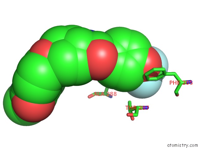
Fluorine Binding Sites:
The binding sites of Fluorine atom in the Crystal Structure of An Epoxide Hydrolase From Trichoderma Reesei in Complex with Inhibitor 4
(pdb code 6n3z). This binding sites where shown within
5.0 Angstroms radius around Fluorine atom.
In total 3 binding sites of Fluorine where determined in the Crystal Structure of An Epoxide Hydrolase From Trichoderma Reesei in Complex with Inhibitor 4, PDB code: 6n3z:
Jump to Fluorine binding site number: 1; 2; 3;
In total 3 binding sites of Fluorine where determined in the Crystal Structure of An Epoxide Hydrolase From Trichoderma Reesei in Complex with Inhibitor 4, PDB code: 6n3z:
Jump to Fluorine binding site number: 1; 2; 3;
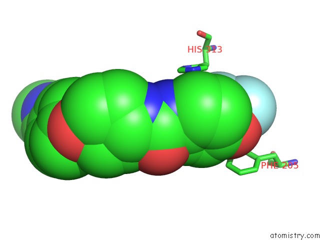
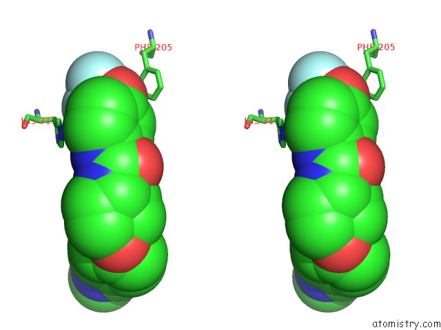
Fluorine binding site 1 out of 3 in 6n3z
Go back to
Fluorine binding site 1 out
of 3 in the Crystal Structure of An Epoxide Hydrolase From Trichoderma Reesei in Complex with Inhibitor 4
Mono view
Stereo pair view
Mono view
Stereo pair view
A full contact list of Fluorine with other atoms in the F binding
site number 1 of Crystal Structure of An Epoxide Hydrolase From Trichoderma Reesei in Complex with Inhibitor 4 within 5.0Å range:
|
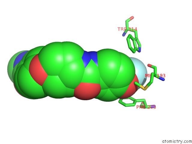
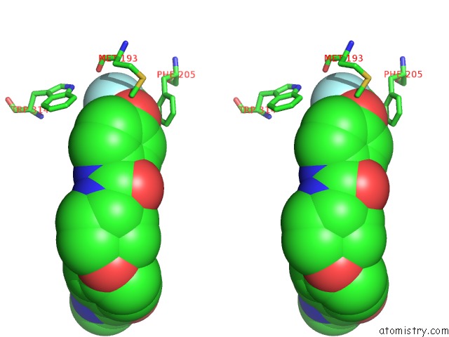
Fluorine binding site 2 out of 3 in 6n3z
Go back to
Fluorine binding site 2 out
of 3 in the Crystal Structure of An Epoxide Hydrolase From Trichoderma Reesei in Complex with Inhibitor 4
Mono view
Stereo pair view
Mono view
Stereo pair view
A full contact list of Fluorine with other atoms in the F binding
site number 2 of Crystal Structure of An Epoxide Hydrolase From Trichoderma Reesei in Complex with Inhibitor 4 within 5.0Å range:
|
Fluorine binding site 3 out of 3 in 6n3z
Go back to
Fluorine binding site 3 out
of 3 in the Crystal Structure of An Epoxide Hydrolase From Trichoderma Reesei in Complex with Inhibitor 4
Mono view

Stereo pair view
Mono view
Stereo pair view
A full contact list of Fluorine with other atoms in the F binding
site number 3 of Crystal Structure of An Epoxide Hydrolase From Trichoderma Reesei in Complex with Inhibitor 4 within 5.0Å range:
|
Reference:
G.S.De Oliveira,
P.P.Adriani,
J.A.Ribeiro,
C.Morisseau,
B.D.Hammock,
M.V.B.Dias,
F.S.Chambergo.
The Molecular Structure of An Epoxide Hydrolase From Trichoderma Reesei in Complex with Urea or Amide-Based Inhibitors. Int. J. Biol. Macromol. V. 129 653 2019.
ISSN: ISSN 1879-0003
PubMed: 30771398
DOI: 10.1016/J.IJBIOMAC.2019.02.070
Page generated: Tue Jul 15 13:17:10 2025
ISSN: ISSN 1879-0003
PubMed: 30771398
DOI: 10.1016/J.IJBIOMAC.2019.02.070
Last articles
Fe in 2YXOFe in 2YRS
Fe in 2YXC
Fe in 2YNM
Fe in 2YVJ
Fe in 2YP1
Fe in 2YU2
Fe in 2YU1
Fe in 2YQB
Fe in 2YOO