Fluorine »
PDB 6ngq-6no8 »
6ngx »
Fluorine in PDB 6ngx: Structure of Rat Neuronal Nitric Oxide Synthase Heme Domain in Complex with 6-(2,3-Difluoro-5-(3-(Methylamino)Prop-1-Yn-1-Yl)Phenethyl)-4- Methylpyridin-2-Amine
Enzymatic activity of Structure of Rat Neuronal Nitric Oxide Synthase Heme Domain in Complex with 6-(2,3-Difluoro-5-(3-(Methylamino)Prop-1-Yn-1-Yl)Phenethyl)-4- Methylpyridin-2-Amine
All present enzymatic activity of Structure of Rat Neuronal Nitric Oxide Synthase Heme Domain in Complex with 6-(2,3-Difluoro-5-(3-(Methylamino)Prop-1-Yn-1-Yl)Phenethyl)-4- Methylpyridin-2-Amine:
1.14.13.39;
1.14.13.39;
Protein crystallography data
The structure of Structure of Rat Neuronal Nitric Oxide Synthase Heme Domain in Complex with 6-(2,3-Difluoro-5-(3-(Methylamino)Prop-1-Yn-1-Yl)Phenethyl)-4- Methylpyridin-2-Amine, PDB code: 6ngx
was solved by
H.Li,
T.L.Poulos,
with X-Ray Crystallography technique. A brief refinement statistics is given in the table below:
| Resolution Low / High (Å) | 39.00 / 1.77 |
| Space group | P 21 21 21 |
| Cell size a, b, c (Å), α, β, γ (°) | 52.024, 111.008, 164.397, 90.00, 90.00, 90.00 |
| R / Rfree (%) | 17.8 / 21.1 |
Other elements in 6ngx:
The structure of Structure of Rat Neuronal Nitric Oxide Synthase Heme Domain in Complex with 6-(2,3-Difluoro-5-(3-(Methylamino)Prop-1-Yn-1-Yl)Phenethyl)-4- Methylpyridin-2-Amine also contains other interesting chemical elements:
| Iron | (Fe) | 2 atoms |
| Zinc | (Zn) | 1 atom |
Fluorine Binding Sites:
The binding sites of Fluorine atom in the Structure of Rat Neuronal Nitric Oxide Synthase Heme Domain in Complex with 6-(2,3-Difluoro-5-(3-(Methylamino)Prop-1-Yn-1-Yl)Phenethyl)-4- Methylpyridin-2-Amine
(pdb code 6ngx). This binding sites where shown within
5.0 Angstroms radius around Fluorine atom.
In total 4 binding sites of Fluorine where determined in the Structure of Rat Neuronal Nitric Oxide Synthase Heme Domain in Complex with 6-(2,3-Difluoro-5-(3-(Methylamino)Prop-1-Yn-1-Yl)Phenethyl)-4- Methylpyridin-2-Amine, PDB code: 6ngx:
Jump to Fluorine binding site number: 1; 2; 3; 4;
In total 4 binding sites of Fluorine where determined in the Structure of Rat Neuronal Nitric Oxide Synthase Heme Domain in Complex with 6-(2,3-Difluoro-5-(3-(Methylamino)Prop-1-Yn-1-Yl)Phenethyl)-4- Methylpyridin-2-Amine, PDB code: 6ngx:
Jump to Fluorine binding site number: 1; 2; 3; 4;
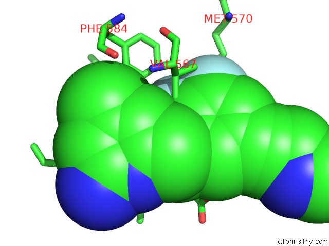
Fluorine binding site 1 out of 4 in 6ngx
Go back to
Fluorine binding site 1 out
of 4 in the Structure of Rat Neuronal Nitric Oxide Synthase Heme Domain in Complex with 6-(2,3-Difluoro-5-(3-(Methylamino)Prop-1-Yn-1-Yl)Phenethyl)-4- Methylpyridin-2-Amine
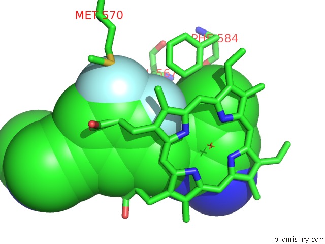
Mono view
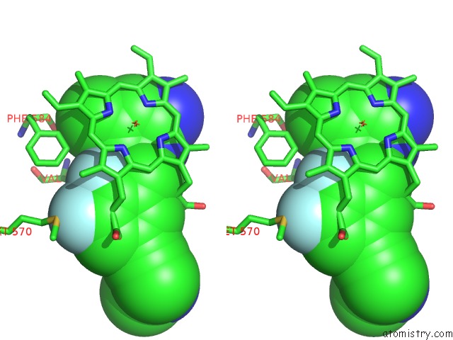
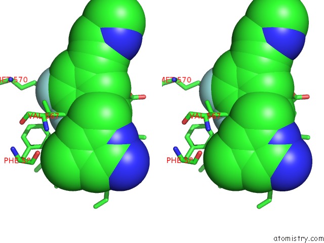
Stereo pair view
Mono view
Stereo pair view
A full contact list of Fluorine with other atoms in the F binding
site number 1 of Structure of Rat Neuronal Nitric Oxide Synthase Heme Domain in Complex with 6-(2,3-Difluoro-5-(3-(Methylamino)Prop-1-Yn-1-Yl)Phenethyl)-4- Methylpyridin-2-Amine within 5.0Å range:
|
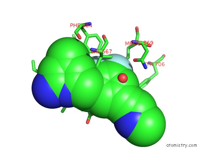
Fluorine binding site 2 out of 4 in 6ngx
Go back to
Fluorine binding site 2 out
of 4 in the Structure of Rat Neuronal Nitric Oxide Synthase Heme Domain in Complex with 6-(2,3-Difluoro-5-(3-(Methylamino)Prop-1-Yn-1-Yl)Phenethyl)-4- Methylpyridin-2-Amine
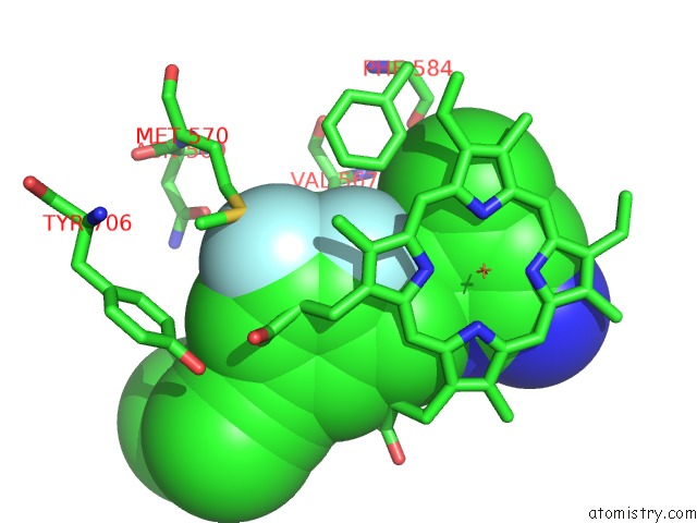
Mono view
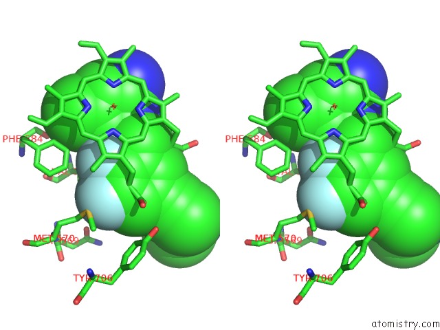
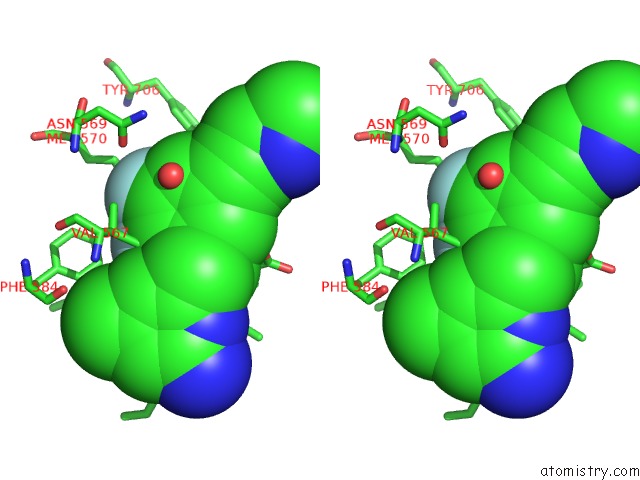
Stereo pair view
Mono view
Stereo pair view
A full contact list of Fluorine with other atoms in the F binding
site number 2 of Structure of Rat Neuronal Nitric Oxide Synthase Heme Domain in Complex with 6-(2,3-Difluoro-5-(3-(Methylamino)Prop-1-Yn-1-Yl)Phenethyl)-4- Methylpyridin-2-Amine within 5.0Å range:
|
Fluorine binding site 3 out of 4 in 6ngx
Go back to
Fluorine binding site 3 out
of 4 in the Structure of Rat Neuronal Nitric Oxide Synthase Heme Domain in Complex with 6-(2,3-Difluoro-5-(3-(Methylamino)Prop-1-Yn-1-Yl)Phenethyl)-4- Methylpyridin-2-Amine
Mono view
Stereo pair view
Mono view
Stereo pair view
A full contact list of Fluorine with other atoms in the F binding
site number 3 of Structure of Rat Neuronal Nitric Oxide Synthase Heme Domain in Complex with 6-(2,3-Difluoro-5-(3-(Methylamino)Prop-1-Yn-1-Yl)Phenethyl)-4- Methylpyridin-2-Amine within 5.0Å range:
|
Fluorine binding site 4 out of 4 in 6ngx
Go back to
Fluorine binding site 4 out
of 4 in the Structure of Rat Neuronal Nitric Oxide Synthase Heme Domain in Complex with 6-(2,3-Difluoro-5-(3-(Methylamino)Prop-1-Yn-1-Yl)Phenethyl)-4- Methylpyridin-2-Amine
Mono view
Stereo pair view
Mono view
Stereo pair view
A full contact list of Fluorine with other atoms in the F binding
site number 4 of Structure of Rat Neuronal Nitric Oxide Synthase Heme Domain in Complex with 6-(2,3-Difluoro-5-(3-(Methylamino)Prop-1-Yn-1-Yl)Phenethyl)-4- Methylpyridin-2-Amine within 5.0Å range:
|
Reference:
H.T.Do,
H.Li,
G.Chreifi,
T.L.Poulos,
R.B.Silverman.
Optimization of Blood-Brain Barrier Permeability with Potent and Selective Human Neuronal Nitric Oxide Synthase Inhibitors Having A 2-Aminopyridine Scaffold. J. Med. Chem. V. 62 2690 2019.
ISSN: ISSN 1520-4804
PubMed: 30802056
DOI: 10.1021/ACS.JMEDCHEM.8B02032
Page generated: Thu Aug 1 22:54:05 2024
ISSN: ISSN 1520-4804
PubMed: 30802056
DOI: 10.1021/ACS.JMEDCHEM.8B02032
Last articles
Zn in 9MJ5Zn in 9HNW
Zn in 9G0L
Zn in 9FNE
Zn in 9DZN
Zn in 9E0I
Zn in 9D32
Zn in 9DAK
Zn in 8ZXC
Zn in 8ZUF