Fluorine »
PDB 6no9-6oab »
6nrg »
Fluorine in PDB 6nrg: Crystal Structure of Human Parp-1 Art Domain Bound to Inhibitor UTT57
Enzymatic activity of Crystal Structure of Human Parp-1 Art Domain Bound to Inhibitor UTT57
All present enzymatic activity of Crystal Structure of Human Parp-1 Art Domain Bound to Inhibitor UTT57:
2.4.2.30;
2.4.2.30;
Protein crystallography data
The structure of Crystal Structure of Human Parp-1 Art Domain Bound to Inhibitor UTT57, PDB code: 6nrg
was solved by
M.F.Langelier,
J.M.Pascal,
with X-Ray Crystallography technique. A brief refinement statistics is given in the table below:
| Resolution Low / High (Å) | 47.72 / 1.70 |
| Space group | I 41 2 2 |
| Cell size a, b, c (Å), α, β, γ (°) | 92.876, 92.876, 138.941, 90.00, 90.00, 90.00 |
| R / Rfree (%) | 15.9 / 18.2 |
Fluorine Binding Sites:
The binding sites of Fluorine atom in the Crystal Structure of Human Parp-1 Art Domain Bound to Inhibitor UTT57
(pdb code 6nrg). This binding sites where shown within
5.0 Angstroms radius around Fluorine atom.
In total only one binding site of Fluorine was determined in the Crystal Structure of Human Parp-1 Art Domain Bound to Inhibitor UTT57, PDB code: 6nrg:
In total only one binding site of Fluorine was determined in the Crystal Structure of Human Parp-1 Art Domain Bound to Inhibitor UTT57, PDB code: 6nrg:
Fluorine binding site 1 out of 1 in 6nrg
Go back to
Fluorine binding site 1 out
of 1 in the Crystal Structure of Human Parp-1 Art Domain Bound to Inhibitor UTT57
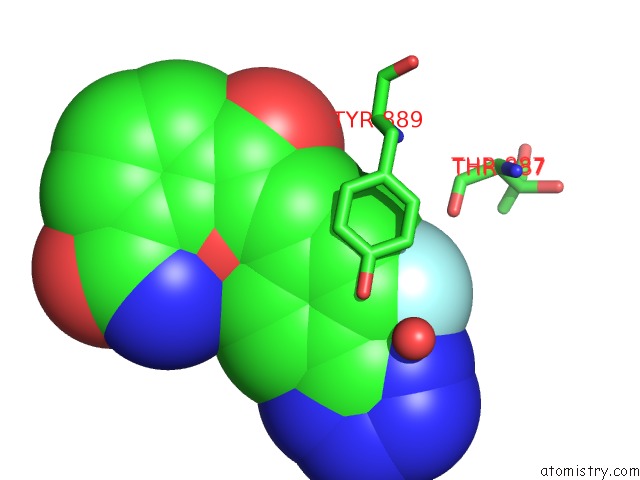
Mono view
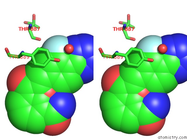
Stereo pair view
Mono view
Stereo pair view
A full contact list of Fluorine with other atoms in the F binding
site number 1 of Crystal Structure of Human Parp-1 Art Domain Bound to Inhibitor UTT57 within 5.0Å range:
|
Reference:
U.K.Velagapudi,
M.F.Langelier,
C.Delgado-Martin,
M.E.Diolaiti,
S.Bakker,
A.Ashworth,
B.A.Patel,
X.Shao,
J.M.Pascal,
T.T.Talele.
Design and Synthesis of Poly(Adp-Ribose) Polymerase Inhibitors: Impact of Adenosine Pocket-Binding Motif Appendage to the 3-Oxo-2,3-Dihydrobenzofuran-7-Carboxamide on Potency and Selectivity. J.Med.Chem. V. 62 5330 2019.
ISSN: ISSN 0022-2623
PubMed: 31042381
DOI: 10.1021/ACS.JMEDCHEM.8B01709
Page generated: Thu Aug 1 23:12:19 2024
ISSN: ISSN 0022-2623
PubMed: 31042381
DOI: 10.1021/ACS.JMEDCHEM.8B01709
Last articles
Ca in 5X7JCa in 5X7P
Ca in 5X7O
Ca in 5X7H
Ca in 5X7G
Ca in 5X6I
Ca in 5X2Q
Ca in 5X2O
Ca in 5X2P
Ca in 5X2M