Fluorine »
PDB 6noj-6oac »
6nwu »
Fluorine in PDB 6nwu: Rorgamma Ligand Binding Domain
Protein crystallography data
The structure of Rorgamma Ligand Binding Domain, PDB code: 6nwu
was solved by
T.S.Strutzenberg,
H.Park,
P.R.Griffin,
with X-Ray Crystallography technique. A brief refinement statistics is given in the table below:
| Resolution Low / High (Å) | 56.19 / 3.20 |
| Space group | P 41 21 2 |
| Cell size a, b, c (Å), α, β, γ (°) | 59.880, 59.880, 162.550, 90.00, 90.00, 90.00 |
| R / Rfree (%) | 21.8 / 27.6 |
Other elements in 6nwu:
The structure of Rorgamma Ligand Binding Domain also contains other interesting chemical elements:
| Chlorine | (Cl) | 2 atoms |
Fluorine Binding Sites:
The binding sites of Fluorine atom in the Rorgamma Ligand Binding Domain
(pdb code 6nwu). This binding sites where shown within
5.0 Angstroms radius around Fluorine atom.
In total 3 binding sites of Fluorine where determined in the Rorgamma Ligand Binding Domain, PDB code: 6nwu:
Jump to Fluorine binding site number: 1; 2; 3;
In total 3 binding sites of Fluorine where determined in the Rorgamma Ligand Binding Domain, PDB code: 6nwu:
Jump to Fluorine binding site number: 1; 2; 3;
Fluorine binding site 1 out of 3 in 6nwu
Go back to
Fluorine binding site 1 out
of 3 in the Rorgamma Ligand Binding Domain
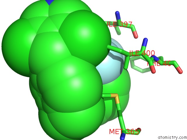
Mono view
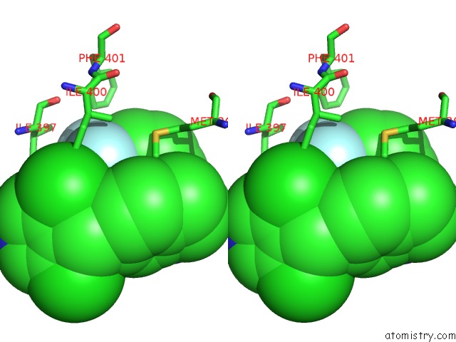
Stereo pair view
Mono view
Stereo pair view
A full contact list of Fluorine with other atoms in the F binding
site number 1 of Rorgamma Ligand Binding Domain within 5.0Å range:
|
Fluorine binding site 2 out of 3 in 6nwu
Go back to
Fluorine binding site 2 out
of 3 in the Rorgamma Ligand Binding Domain
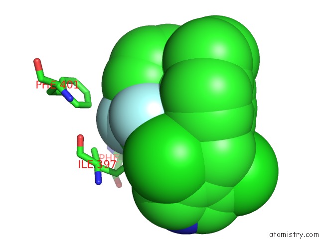
Mono view

Stereo pair view
Mono view
Stereo pair view
A full contact list of Fluorine with other atoms in the F binding
site number 2 of Rorgamma Ligand Binding Domain within 5.0Å range:
|
Fluorine binding site 3 out of 3 in 6nwu
Go back to
Fluorine binding site 3 out
of 3 in the Rorgamma Ligand Binding Domain
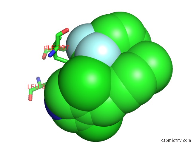
Mono view
Stereo pair view
Mono view
Stereo pair view
A full contact list of Fluorine with other atoms in the F binding
site number 3 of Rorgamma Ligand Binding Domain within 5.0Å range:
|
Reference:
T.S.Strutzenberg,
R.D.Garcia-Ordonez,
S.J.Novick,
H.Park,
M.R.Chang,
C.Doebellin,
Y.He,
R.Patouret,
T.M.Kamenecka,
P.R.Griffin.
Hdx-Ms Reveals Structural Determinants For Ror Gamma Hyperactivation By Synthetic Agonists. Elife V. 8 2019.
ISSN: ESSN 2050-084X
PubMed: 31172947
DOI: 10.7554/ELIFE.47172
Page generated: Tue Jul 15 13:52:29 2025
ISSN: ESSN 2050-084X
PubMed: 31172947
DOI: 10.7554/ELIFE.47172
Last articles
Fe in 2YXOFe in 2YRS
Fe in 2YXC
Fe in 2YNM
Fe in 2YVJ
Fe in 2YP1
Fe in 2YU2
Fe in 2YU1
Fe in 2YQB
Fe in 2YOO