Fluorine »
PDB 6q0t-6qhv »
6qdy »
Fluorine in PDB 6qdy: The Crystal Structure of Sporosarcina Pasteurii Urease in Complex with Its Substrate Urea
Enzymatic activity of The Crystal Structure of Sporosarcina Pasteurii Urease in Complex with Its Substrate Urea
All present enzymatic activity of The Crystal Structure of Sporosarcina Pasteurii Urease in Complex with Its Substrate Urea:
3.5.1.5;
3.5.1.5;
Protein crystallography data
The structure of The Crystal Structure of Sporosarcina Pasteurii Urease in Complex with Its Substrate Urea, PDB code: 6qdy
was solved by
L.Mazzei,
M.Cianci,
S.Benini,
S.Ciurli,
with X-Ray Crystallography technique. A brief refinement statistics is given in the table below:
| Resolution Low / High (Å) | 97.54 / 1.42 |
| Space group | P 63 2 2 |
| Cell size a, b, c (Å), α, β, γ (°) | 131.565, 131.565, 188.683, 90.00, 90.00, 120.00 |
| R / Rfree (%) | 9.5 / 11.9 |
Other elements in 6qdy:
The structure of The Crystal Structure of Sporosarcina Pasteurii Urease in Complex with Its Substrate Urea also contains other interesting chemical elements:
| Nickel | (Ni) | 2 atoms |
Fluorine Binding Sites:
The binding sites of Fluorine atom in the The Crystal Structure of Sporosarcina Pasteurii Urease in Complex with Its Substrate Urea
(pdb code 6qdy). This binding sites where shown within
5.0 Angstroms radius around Fluorine atom.
In total only one binding site of Fluorine was determined in the The Crystal Structure of Sporosarcina Pasteurii Urease in Complex with Its Substrate Urea, PDB code: 6qdy:
In total only one binding site of Fluorine was determined in the The Crystal Structure of Sporosarcina Pasteurii Urease in Complex with Its Substrate Urea, PDB code: 6qdy:
Fluorine binding site 1 out of 1 in 6qdy
Go back to
Fluorine binding site 1 out
of 1 in the The Crystal Structure of Sporosarcina Pasteurii Urease in Complex with Its Substrate Urea
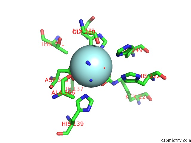
Mono view
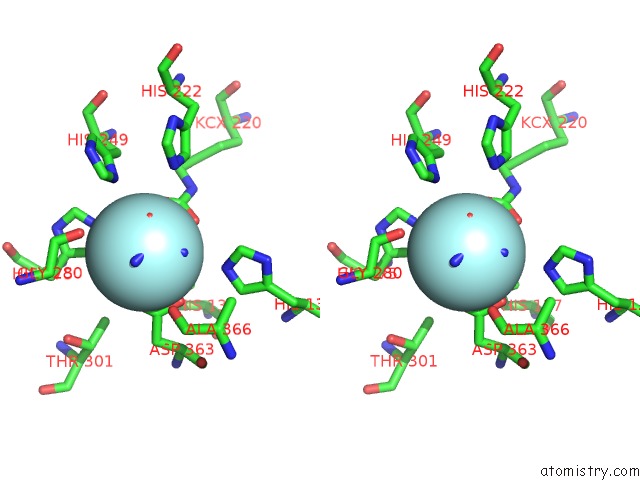
Stereo pair view
Mono view
Stereo pair view
A full contact list of Fluorine with other atoms in the F binding
site number 1 of The Crystal Structure of Sporosarcina Pasteurii Urease in Complex with Its Substrate Urea within 5.0Å range:
|
Reference:
L.Mazzei,
M.Cianci,
S.Benini,
S.Ciurli.
The Structure of the Elusive Urease-Urea Complex Unveils the Mechanism of A Paradigmatic Nickel-Dependent Enzyme. Angew.Chem.Int.Ed.Engl. V. 58 7415 2019.
ISSN: ESSN 1521-3773
PubMed: 30969470
DOI: 10.1002/ANIE.201903565
Page generated: Fri Aug 2 00:46:53 2024
ISSN: ESSN 1521-3773
PubMed: 30969470
DOI: 10.1002/ANIE.201903565
Last articles
Zn in 9J0NZn in 9J0O
Zn in 9J0P
Zn in 9FJX
Zn in 9EKB
Zn in 9C0F
Zn in 9CAH
Zn in 9CH0
Zn in 9CH3
Zn in 9CH1