Fluorine »
PDB 6qy8-6rkn »
6r0h »
Fluorine in PDB 6r0h: Glycogen Phosphorylase B in Complex with 3
Enzymatic activity of Glycogen Phosphorylase B in Complex with 3
All present enzymatic activity of Glycogen Phosphorylase B in Complex with 3:
2.4.1.1;
2.4.1.1;
Protein crystallography data
The structure of Glycogen Phosphorylase B in Complex with 3, PDB code: 6r0h
was solved by
S.A.Tsagkarakou,
M.S.Koulas,
E.Kyriakis,
G.A.Stravodimos,
V.T.Skamnaki,
D.D.Leonidas,
with X-Ray Crystallography technique. A brief refinement statistics is given in the table below:
| Resolution Low / High (Å) | 13.71 / 2.50 |
| Space group | P 43 21 2 |
| Cell size a, b, c (Å), α, β, γ (°) | 128.287, 128.287, 116.120, 90.00, 90.00, 90.00 |
| R / Rfree (%) | 15.9 / 21.3 |
Fluorine Binding Sites:
The binding sites of Fluorine atom in the Glycogen Phosphorylase B in Complex with 3
(pdb code 6r0h). This binding sites where shown within
5.0 Angstroms radius around Fluorine atom.
In total only one binding site of Fluorine was determined in the Glycogen Phosphorylase B in Complex with 3, PDB code: 6r0h:
In total only one binding site of Fluorine was determined in the Glycogen Phosphorylase B in Complex with 3, PDB code: 6r0h:
Fluorine binding site 1 out of 1 in 6r0h
Go back to
Fluorine binding site 1 out
of 1 in the Glycogen Phosphorylase B in Complex with 3
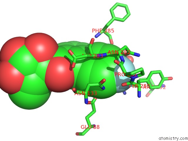
Mono view
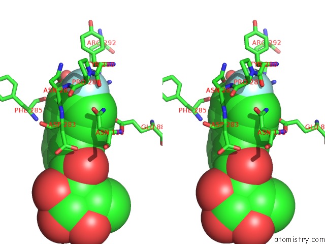
Stereo pair view
Mono view
Stereo pair view
A full contact list of Fluorine with other atoms in the F binding
site number 1 of Glycogen Phosphorylase B in Complex with 3 within 5.0Å range:
|
Reference:
T.Fischer,
S.M.Koulas,
A.S.Tsagkarakou,
E.Kyriakis,
G.A.Stravodimos,
V.T.Skamnaki,
P.G.V.Liggri,
S.E.Zographos,
R.Riedl,
D.D.Leonidas.
High Consistency of Structure-Based Design and X-Ray Crystallography: Design, Synthesis, Kinetic Evaluation and Crystallographic Binding Mode Determination of Biphenyl-N-Acyl-Beta-D-Glucopyranosylamines As Glycogen Phosphorylase Inhibitors. Molecules V. 24 2019.
ISSN: ESSN 1420-3049
PubMed: 30987252
DOI: 10.3390/MOLECULES24071322
Page generated: Fri Aug 2 01:06:18 2024
ISSN: ESSN 1420-3049
PubMed: 30987252
DOI: 10.3390/MOLECULES24071322
Last articles
Zn in 9MJ5Zn in 9HNW
Zn in 9G0L
Zn in 9FNE
Zn in 9DZN
Zn in 9E0I
Zn in 9D32
Zn in 9DAK
Zn in 8ZXC
Zn in 8ZUF