Fluorine »
PDB 6qy8-6rkn »
6rj7 »
Fluorine in PDB 6rj7: Crystal Structure of the 19F Labelled Oxa-48
Enzymatic activity of Crystal Structure of the 19F Labelled Oxa-48
All present enzymatic activity of Crystal Structure of the 19F Labelled Oxa-48:
3.5.2.6;
3.5.2.6;
Protein crystallography data
The structure of Crystal Structure of the 19F Labelled Oxa-48, PDB code: 6rj7
was solved by
J.Brem,
C.Lohans,
C.Schofield,
with X-Ray Crystallography technique. A brief refinement statistics is given in the table below:
| Resolution Low / High (Å) | 66.40 / 1.73 |
| Space group | P 1 21 1 |
| Cell size a, b, c (Å), α, β, γ (°) | 64.360, 58.030, 66.430, 90.00, 91.49, 90.00 |
| R / Rfree (%) | 17.7 / 21 |
Other elements in 6rj7:
The structure of Crystal Structure of the 19F Labelled Oxa-48 also contains other interesting chemical elements:
| Calcium | (Ca) | 1 atom |
| Chlorine | (Cl) | 1 atom |
Fluorine Binding Sites:
The binding sites of Fluorine atom in the Crystal Structure of the 19F Labelled Oxa-48
(pdb code 6rj7). This binding sites where shown within
5.0 Angstroms radius around Fluorine atom.
In total 6 binding sites of Fluorine where determined in the Crystal Structure of the 19F Labelled Oxa-48, PDB code: 6rj7:
Jump to Fluorine binding site number: 1; 2; 3; 4; 5; 6;
In total 6 binding sites of Fluorine where determined in the Crystal Structure of the 19F Labelled Oxa-48, PDB code: 6rj7:
Jump to Fluorine binding site number: 1; 2; 3; 4; 5; 6;
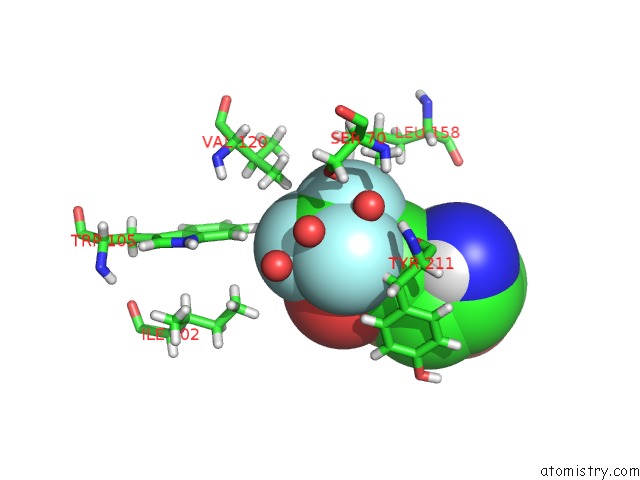
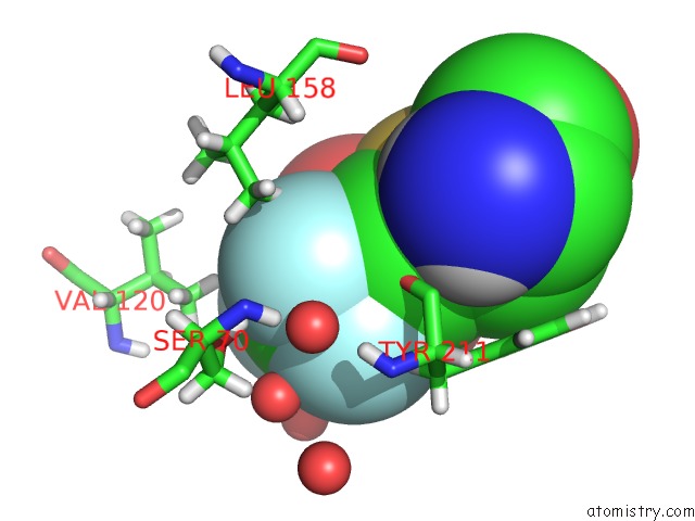
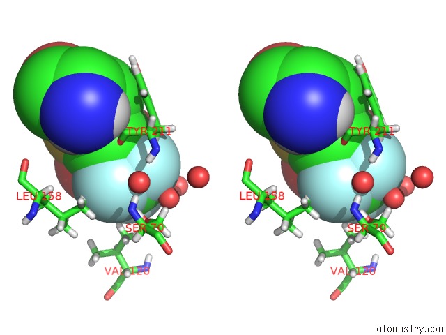
Fluorine binding site 1 out of 6 in 6rj7
Go back to
Fluorine binding site 1 out
of 6 in the Crystal Structure of the 19F Labelled Oxa-48
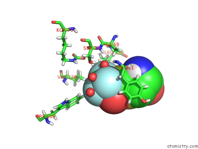
Mono view
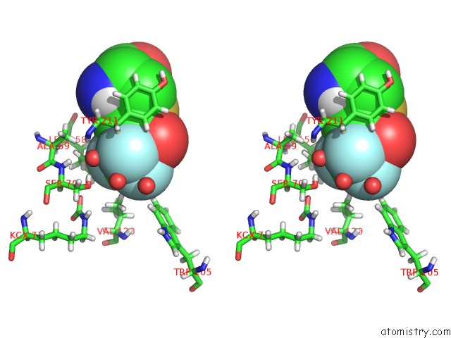
Stereo pair view
Mono view
Stereo pair view
A full contact list of Fluorine with other atoms in the F binding
site number 1 of Crystal Structure of the 19F Labelled Oxa-48 within 5.0Å range:
|
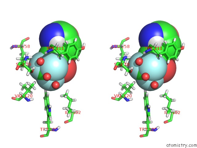
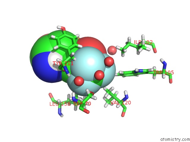
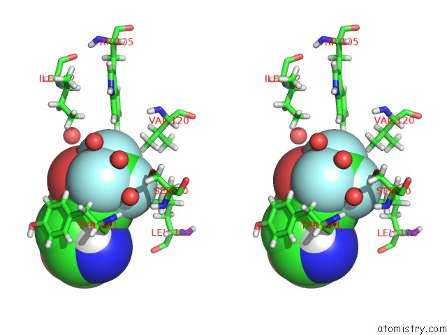
Fluorine binding site 2 out of 6 in 6rj7
Go back to
Fluorine binding site 2 out
of 6 in the Crystal Structure of the 19F Labelled Oxa-48
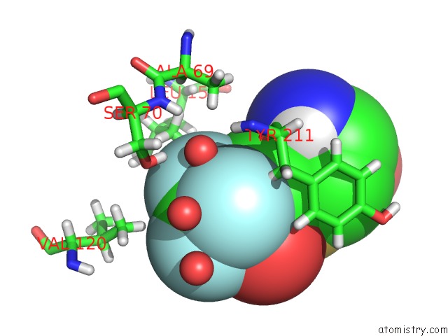
Mono view
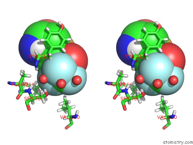
Stereo pair view
Mono view
Stereo pair view
A full contact list of Fluorine with other atoms in the F binding
site number 2 of Crystal Structure of the 19F Labelled Oxa-48 within 5.0Å range:
|
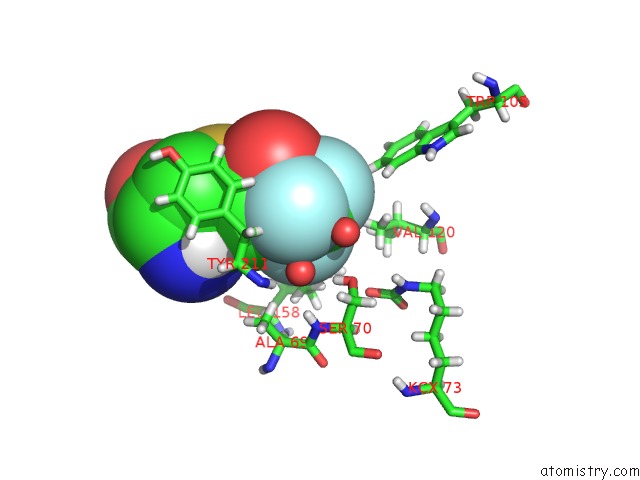
Fluorine binding site 3 out of 6 in 6rj7
Go back to
Fluorine binding site 3 out
of 6 in the Crystal Structure of the 19F Labelled Oxa-48
Mono view
Stereo pair view
Mono view
Stereo pair view
A full contact list of Fluorine with other atoms in the F binding
site number 3 of Crystal Structure of the 19F Labelled Oxa-48 within 5.0Å range:
|
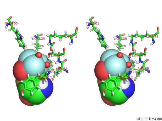
Fluorine binding site 4 out of 6 in 6rj7
Go back to
Fluorine binding site 4 out
of 6 in the Crystal Structure of the 19F Labelled Oxa-48
Mono view
Stereo pair view
Mono view
Stereo pair view
A full contact list of Fluorine with other atoms in the F binding
site number 4 of Crystal Structure of the 19F Labelled Oxa-48 within 5.0Å range:
|
Fluorine binding site 5 out of 6 in 6rj7
Go back to
Fluorine binding site 5 out
of 6 in the Crystal Structure of the 19F Labelled Oxa-48
Mono view
Stereo pair view
Mono view
Stereo pair view
A full contact list of Fluorine with other atoms in the F binding
site number 5 of Crystal Structure of the 19F Labelled Oxa-48 within 5.0Å range:
|
Fluorine binding site 6 out of 6 in 6rj7
Go back to
Fluorine binding site 6 out
of 6 in the Crystal Structure of the 19F Labelled Oxa-48
Mono view
Stereo pair view
Mono view
Stereo pair view
A full contact list of Fluorine with other atoms in the F binding
site number 6 of Crystal Structure of the 19F Labelled Oxa-48 within 5.0Å range:
|
Reference:
E.Van Groesen,
C.T.Lohans,
J.Brem,
K.M.J.Aertker,
T.D.W.Claridge,
C.J.Schofield.
19F uc(Nmr) Monitoring of Reversible Protein Post-Translational Modifications: Class D Beta-Lactamase Carbamylation and Inhibition. Chemistry V. 25 11837 2019.
ISSN: ISSN 0947-6539
PubMed: 31310409
DOI: 10.1002/CHEM.201902529
Page generated: Fri Aug 2 01:15:12 2024
ISSN: ISSN 0947-6539
PubMed: 31310409
DOI: 10.1002/CHEM.201902529
Last articles
F in 4G8OF in 4G6O
F in 4G2R
F in 4G5P
F in 4G5J
F in 4G3G
F in 4G3F
F in 4G2I
F in 4G2H
F in 4G31