Fluorine »
PDB 6rkp-6rzg »
6rv3 »
Fluorine in PDB 6rv3: Crystal Structure of the Human Two Pore Domain Potassium Ion Channel Task-1 (K2P3.1) in A Closed Conformation with A Bound Inhibitor Bay 1000493
Protein crystallography data
The structure of Crystal Structure of the Human Two Pore Domain Potassium Ion Channel Task-1 (K2P3.1) in A Closed Conformation with A Bound Inhibitor Bay 1000493, PDB code: 6rv3
was solved by
K.E.J.Rodstrom,
A.C.W.Pike,
W.Zhang,
A.Quigley,
D.Speedman,
S.M.M.Mukhopadhyay,
L.Shrestha,
R.Chalk,
S.Venkaya,
S.R.Bushell,
A.Tessitore,
N.Burgess-Brown,
C.H.Arrowsmith,
A.M.Edwards,
C.Bountra,
E.P.Carpenter,
Structural Genomics Consortium (Sgc),
with X-Ray Crystallography technique. A brief refinement statistics is given in the table below:
| Resolution Low / High (Å) | 41.31 / 2.90 |
| Space group | P 2 21 21 |
| Cell size a, b, c (Å), α, β, γ (°) | 45.070, 204.500, 239.320, 90.00, 90.00, 90.00 |
| R / Rfree (%) | 23.5 / 24.2 |
Other elements in 6rv3:
The structure of Crystal Structure of the Human Two Pore Domain Potassium Ion Channel Task-1 (K2P3.1) in A Closed Conformation with A Bound Inhibitor Bay 1000493 also contains other interesting chemical elements:
| Bromine | (Br) | 4 atoms |
| Potassium | (K) | 8 atoms |
Fluorine Binding Sites:
The binding sites of Fluorine atom in the Crystal Structure of the Human Two Pore Domain Potassium Ion Channel Task-1 (K2P3.1) in A Closed Conformation with A Bound Inhibitor Bay 1000493
(pdb code 6rv3). This binding sites where shown within
5.0 Angstroms radius around Fluorine atom.
In total 4 binding sites of Fluorine where determined in the Crystal Structure of the Human Two Pore Domain Potassium Ion Channel Task-1 (K2P3.1) in A Closed Conformation with A Bound Inhibitor Bay 1000493, PDB code: 6rv3:
Jump to Fluorine binding site number: 1; 2; 3; 4;
In total 4 binding sites of Fluorine where determined in the Crystal Structure of the Human Two Pore Domain Potassium Ion Channel Task-1 (K2P3.1) in A Closed Conformation with A Bound Inhibitor Bay 1000493, PDB code: 6rv3:
Jump to Fluorine binding site number: 1; 2; 3; 4;
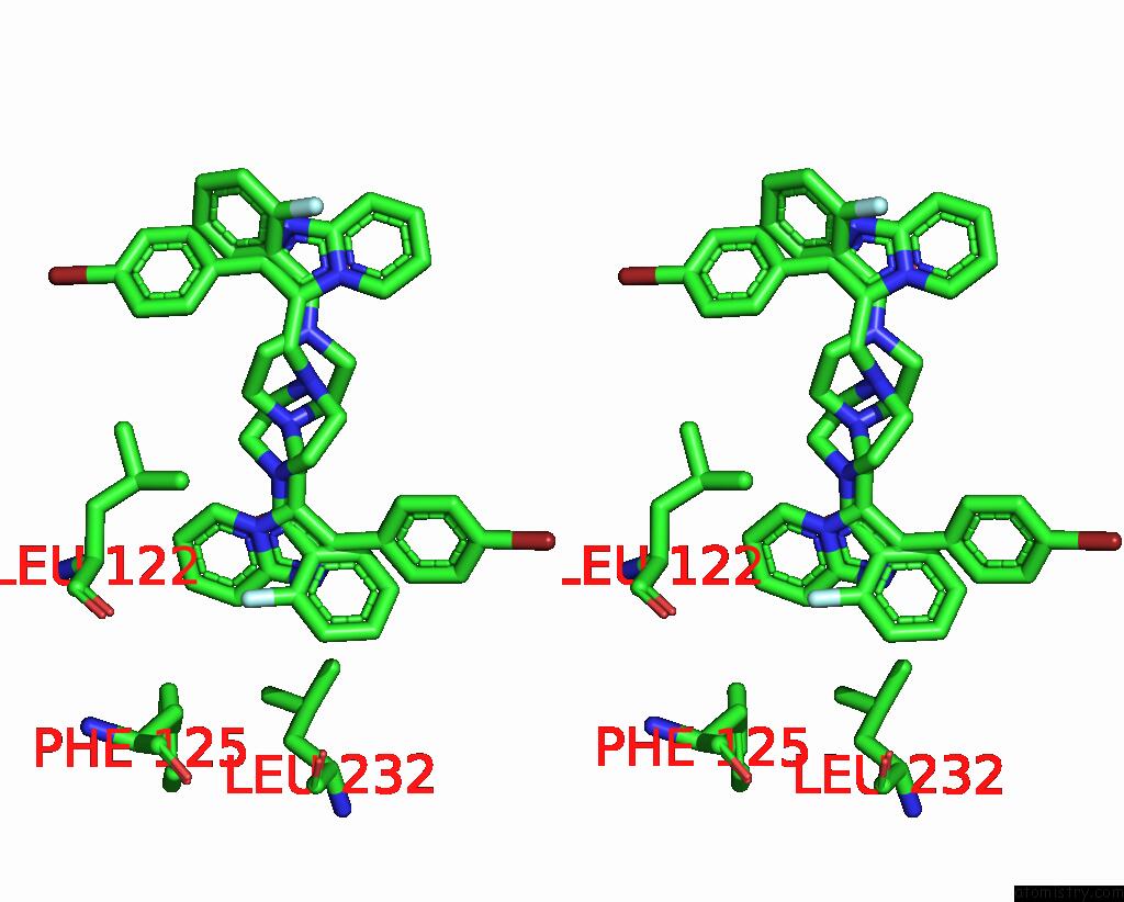
Fluorine binding site 1 out of 4 in 6rv3
Go back to
Fluorine binding site 1 out
of 4 in the Crystal Structure of the Human Two Pore Domain Potassium Ion Channel Task-1 (K2P3.1) in A Closed Conformation with A Bound Inhibitor Bay 1000493
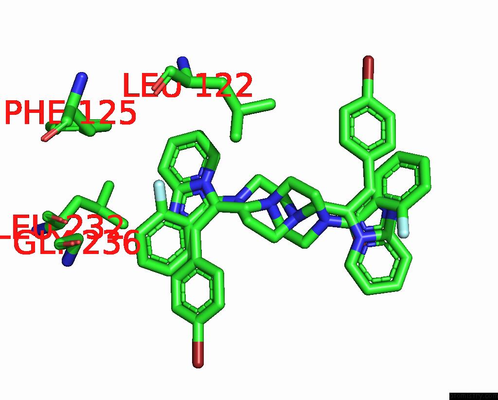
Mono view

Stereo pair view
Mono view
Stereo pair view
A full contact list of Fluorine with other atoms in the F binding
site number 1 of Crystal Structure of the Human Two Pore Domain Potassium Ion Channel Task-1 (K2P3.1) in A Closed Conformation with A Bound Inhibitor Bay 1000493 within 5.0Å range:
|
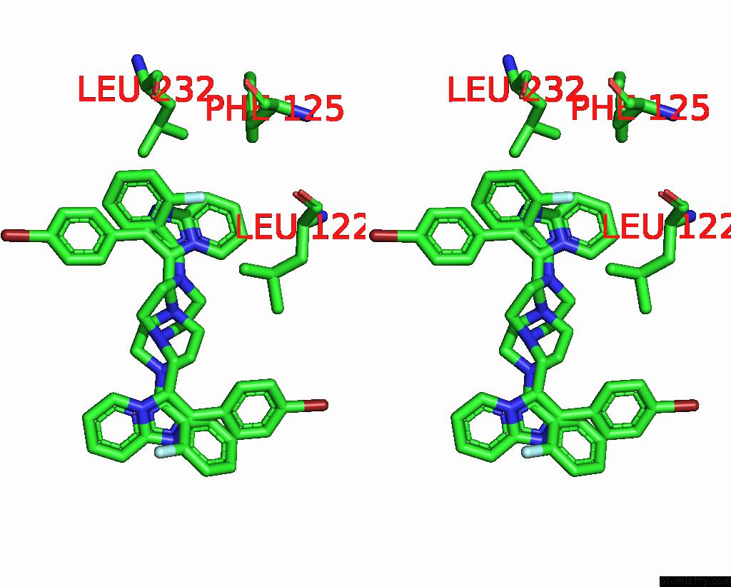
Fluorine binding site 2 out of 4 in 6rv3
Go back to
Fluorine binding site 2 out
of 4 in the Crystal Structure of the Human Two Pore Domain Potassium Ion Channel Task-1 (K2P3.1) in A Closed Conformation with A Bound Inhibitor Bay 1000493
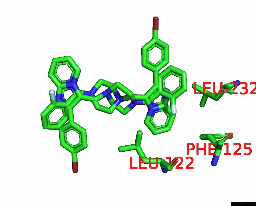
Mono view
Stereo pair view
Mono view
Stereo pair view
A full contact list of Fluorine with other atoms in the F binding
site number 2 of Crystal Structure of the Human Two Pore Domain Potassium Ion Channel Task-1 (K2P3.1) in A Closed Conformation with A Bound Inhibitor Bay 1000493 within 5.0Å range:
|
Fluorine binding site 3 out of 4 in 6rv3
Go back to
Fluorine binding site 3 out
of 4 in the Crystal Structure of the Human Two Pore Domain Potassium Ion Channel Task-1 (K2P3.1) in A Closed Conformation with A Bound Inhibitor Bay 1000493
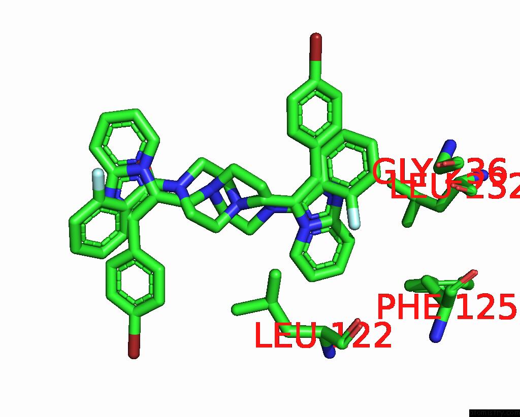
Mono view

Stereo pair view
Mono view
Stereo pair view
A full contact list of Fluorine with other atoms in the F binding
site number 3 of Crystal Structure of the Human Two Pore Domain Potassium Ion Channel Task-1 (K2P3.1) in A Closed Conformation with A Bound Inhibitor Bay 1000493 within 5.0Å range:
|
Fluorine binding site 4 out of 4 in 6rv3
Go back to
Fluorine binding site 4 out
of 4 in the Crystal Structure of the Human Two Pore Domain Potassium Ion Channel Task-1 (K2P3.1) in A Closed Conformation with A Bound Inhibitor Bay 1000493

Mono view
Stereo pair view
Mono view
Stereo pair view
A full contact list of Fluorine with other atoms in the F binding
site number 4 of Crystal Structure of the Human Two Pore Domain Potassium Ion Channel Task-1 (K2P3.1) in A Closed Conformation with A Bound Inhibitor Bay 1000493 within 5.0Å range:
|
Reference:
K.E.J.Rodstrom,
A.K.Kiper,
W.Zhang,
S.Rinne,
A.C.W.Pike,
M.Goldstein,
L.Conrad,
M.Delbeck,
M.Hahn,
H.Meier,
M.Platzk,
A.Quigley,
D.Speedman,
L.Shrestha,
S.M.M.Mukhopadhyay,
N.A.Burgess-Brown,
S.J.Tucker,
T.Mueller,
N.Decher,
E.P.Carpenter.
A Unique Lower X-Gate in Task Channels Sequesters Inhibitors Within the Vestibule To Be Published.
Page generated: Fri Aug 2 01:25:16 2024
Last articles
Zn in 9J0NZn in 9J0O
Zn in 9J0P
Zn in 9FJX
Zn in 9EKB
Zn in 9C0F
Zn in 9CAH
Zn in 9CH0
Zn in 9CH3
Zn in 9CH1