Fluorine »
PDB 6slz-6tan »
6sov »
Fluorine in PDB 6sov: Fragments KCL_615 and KCL_802 in Complex with Map Kinase P38-Alpha
Enzymatic activity of Fragments KCL_615 and KCL_802 in Complex with Map Kinase P38-Alpha
All present enzymatic activity of Fragments KCL_615 and KCL_802 in Complex with Map Kinase P38-Alpha:
2.7.11.24;
2.7.11.24;
Protein crystallography data
The structure of Fragments KCL_615 and KCL_802 in Complex with Map Kinase P38-Alpha, PDB code: 6sov
was solved by
C.E.Nichols,
G.F.De Nicola,
with X-Ray Crystallography technique. A brief refinement statistics is given in the table below:
| Resolution Low / High (Å) | 63.54 / 1.31 |
| Space group | I 2 2 2 |
| Cell size a, b, c (Å), α, β, γ (°) | 80.893, 102.685, 103.687, 90.00, 90.00, 90.00 |
| R / Rfree (%) | 22.1 / 23.7 |
Other elements in 6sov:
The structure of Fragments KCL_615 and KCL_802 in Complex with Map Kinase P38-Alpha also contains other interesting chemical elements:
| Calcium | (Ca) | 4 atoms |
Fluorine Binding Sites:
The binding sites of Fluorine atom in the Fragments KCL_615 and KCL_802 in Complex with Map Kinase P38-Alpha
(pdb code 6sov). This binding sites where shown within
5.0 Angstroms radius around Fluorine atom.
In total only one binding site of Fluorine was determined in the Fragments KCL_615 and KCL_802 in Complex with Map Kinase P38-Alpha, PDB code: 6sov:
In total only one binding site of Fluorine was determined in the Fragments KCL_615 and KCL_802 in Complex with Map Kinase P38-Alpha, PDB code: 6sov:
Fluorine binding site 1 out of 1 in 6sov
Go back to
Fluorine binding site 1 out
of 1 in the Fragments KCL_615 and KCL_802 in Complex with Map Kinase P38-Alpha
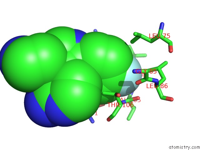
Mono view
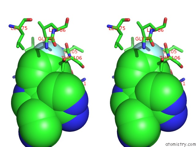
Stereo pair view
Mono view
Stereo pair view
A full contact list of Fluorine with other atoms in the F binding
site number 1 of Fragments KCL_615 and KCL_802 in Complex with Map Kinase P38-Alpha within 5.0Å range:
|
Reference:
G.F.De Nicola,
C.E.Nichols.
Targeting the P38 / TAB1 Interface By Single Fragment in-Crystal Screening and Structure-Based Drug Design. To Be Published.
Page generated: Fri Aug 2 01:48:07 2024
Last articles
Zn in 9J0NZn in 9J0O
Zn in 9J0P
Zn in 9FJX
Zn in 9EKB
Zn in 9C0F
Zn in 9CAH
Zn in 9CH0
Zn in 9CH3
Zn in 9CH1