Fluorine »
PDB 6slz-6tan »
6sq5 »
Fluorine in PDB 6sq5: Crystal Structure of M. Tuberculosis Inha in Complex with Nad+ and 3- [3-(Trifluoromethyl)Phenyl]Prop-2-Enoic Acid
Enzymatic activity of Crystal Structure of M. Tuberculosis Inha in Complex with Nad+ and 3- [3-(Trifluoromethyl)Phenyl]Prop-2-Enoic Acid
All present enzymatic activity of Crystal Structure of M. Tuberculosis Inha in Complex with Nad+ and 3- [3-(Trifluoromethyl)Phenyl]Prop-2-Enoic Acid:
1.3.1.9;
1.3.1.9;
Protein crystallography data
The structure of Crystal Structure of M. Tuberculosis Inha in Complex with Nad+ and 3- [3-(Trifluoromethyl)Phenyl]Prop-2-Enoic Acid, PDB code: 6sq5
was solved by
V.Mendes,
M.Sabbah,
A.G.Coyne,
C.Abell,
T.L.Blundell,
with X-Ray Crystallography technique. A brief refinement statistics is given in the table below:
| Resolution Low / High (Å) | 72.35 / 1.84 |
| Space group | P 62 2 2 |
| Cell size a, b, c (Å), α, β, γ (°) | 97.533, 97.533, 140.229, 90.00, 90.00, 120.00 |
| R / Rfree (%) | 16.3 / 18.2 |
Fluorine Binding Sites:
The binding sites of Fluorine atom in the Crystal Structure of M. Tuberculosis Inha in Complex with Nad+ and 3- [3-(Trifluoromethyl)Phenyl]Prop-2-Enoic Acid
(pdb code 6sq5). This binding sites where shown within
5.0 Angstroms radius around Fluorine atom.
In total 3 binding sites of Fluorine where determined in the Crystal Structure of M. Tuberculosis Inha in Complex with Nad+ and 3- [3-(Trifluoromethyl)Phenyl]Prop-2-Enoic Acid, PDB code: 6sq5:
Jump to Fluorine binding site number: 1; 2; 3;
In total 3 binding sites of Fluorine where determined in the Crystal Structure of M. Tuberculosis Inha in Complex with Nad+ and 3- [3-(Trifluoromethyl)Phenyl]Prop-2-Enoic Acid, PDB code: 6sq5:
Jump to Fluorine binding site number: 1; 2; 3;
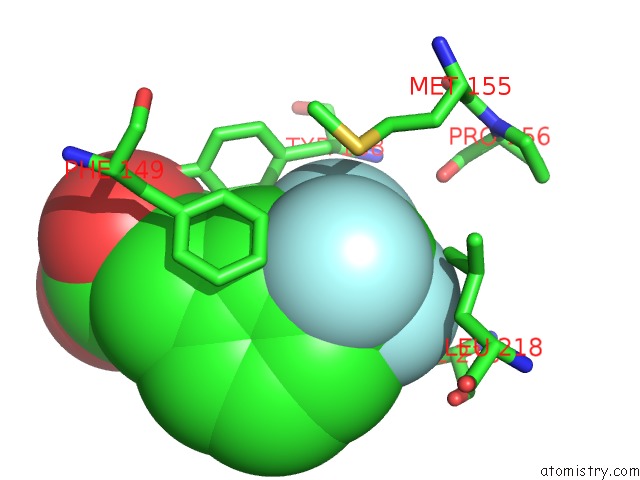
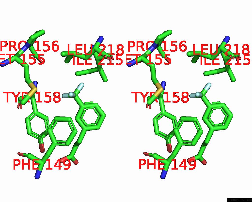
Fluorine binding site 1 out of 3 in 6sq5
Go back to
Fluorine binding site 1 out
of 3 in the Crystal Structure of M. Tuberculosis Inha in Complex with Nad+ and 3- [3-(Trifluoromethyl)Phenyl]Prop-2-Enoic Acid
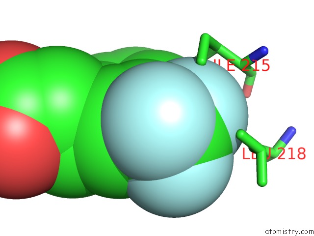
Mono view
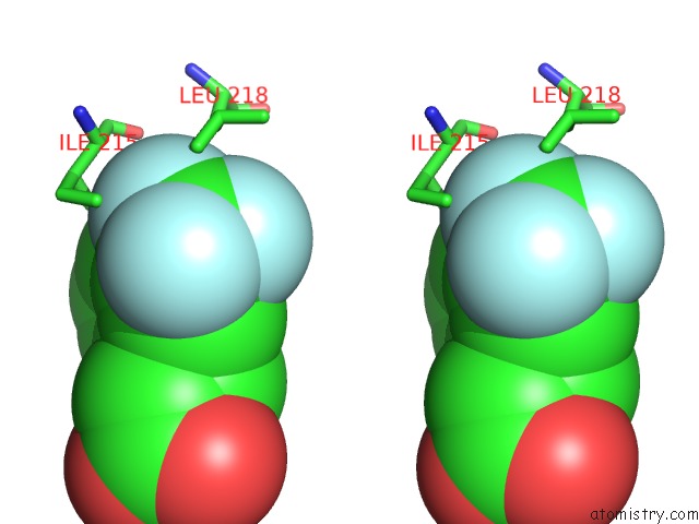
Stereo pair view
Mono view
Stereo pair view
A full contact list of Fluorine with other atoms in the F binding
site number 1 of Crystal Structure of M. Tuberculosis Inha in Complex with Nad+ and 3- [3-(Trifluoromethyl)Phenyl]Prop-2-Enoic Acid within 5.0Å range:
|
Fluorine binding site 2 out of 3 in 6sq5
Go back to
Fluorine binding site 2 out
of 3 in the Crystal Structure of M. Tuberculosis Inha in Complex with Nad+ and 3- [3-(Trifluoromethyl)Phenyl]Prop-2-Enoic Acid
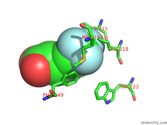
Mono view
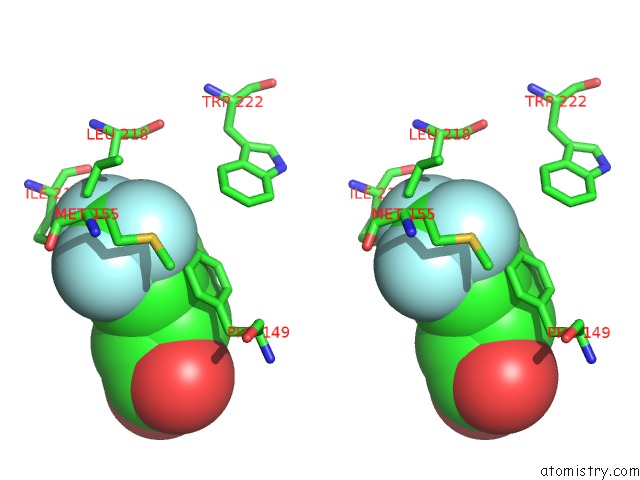
Stereo pair view
Mono view
Stereo pair view
A full contact list of Fluorine with other atoms in the F binding
site number 2 of Crystal Structure of M. Tuberculosis Inha in Complex with Nad+ and 3- [3-(Trifluoromethyl)Phenyl]Prop-2-Enoic Acid within 5.0Å range:
|
Fluorine binding site 3 out of 3 in 6sq5
Go back to
Fluorine binding site 3 out
of 3 in the Crystal Structure of M. Tuberculosis Inha in Complex with Nad+ and 3- [3-(Trifluoromethyl)Phenyl]Prop-2-Enoic Acid
Mono view
Stereo pair view
Mono view
Stereo pair view
A full contact list of Fluorine with other atoms in the F binding
site number 3 of Crystal Structure of M. Tuberculosis Inha in Complex with Nad+ and 3- [3-(Trifluoromethyl)Phenyl]Prop-2-Enoic Acid within 5.0Å range:
|
Reference:
M.Sabbah,
V.Mendes,
R.G.Vistal,
D.M.Dias,
M.Zahorszka,
K.Mikusova,
J.Kordulakova,
A.G.Coyne,
T.L.Blundell,
C.Abell.
Fragment-Based Design of Mycobacterium Tuberculosis Inha Inhibitors. J.Med.Chem. 2020.
ISSN: ISSN 0022-2623
PubMed: 32240584
DOI: 10.1021/ACS.JMEDCHEM.0C00007
Page generated: Fri Aug 2 01:48:07 2024
ISSN: ISSN 0022-2623
PubMed: 32240584
DOI: 10.1021/ACS.JMEDCHEM.0C00007
Last articles
F in 4G6OF in 4G2R
F in 4G5P
F in 4G5J
F in 4G3G
F in 4G3F
F in 4G2I
F in 4G2H
F in 4G31
F in 4G1W