Fluorine »
PDB 6tvl-6ugr »
6udm »
Fluorine in PDB 6udm: Structure of Human Cytochrome P450 1A1 with Duocarmycin Prodrug (S) Ict-2726
Enzymatic activity of Structure of Human Cytochrome P450 1A1 with Duocarmycin Prodrug (S) Ict-2726
All present enzymatic activity of Structure of Human Cytochrome P450 1A1 with Duocarmycin Prodrug (S) Ict-2726:
1.14.14.1; 4.2.1.152;
1.14.14.1; 4.2.1.152;
Protein crystallography data
The structure of Structure of Human Cytochrome P450 1A1 with Duocarmycin Prodrug (S) Ict-2726, PDB code: 6udm
was solved by
A.G.Bart,
E.E.Scott,
with X-Ray Crystallography technique. A brief refinement statistics is given in the table below:
| Resolution Low / High (Å) | 39.64 / 3.08 |
| Space group | P 31 2 1 |
| Cell size a, b, c (Å), α, β, γ (°) | 241.281, 241.281, 125.298, 90.00, 90.00, 120.00 |
| R / Rfree (%) | 20.9 / 22.1 |
Other elements in 6udm:
The structure of Structure of Human Cytochrome P450 1A1 with Duocarmycin Prodrug (S) Ict-2726 also contains other interesting chemical elements:
| Iron | (Fe) | 4 atoms |
| Chlorine | (Cl) | 4 atoms |
Fluorine Binding Sites:
The binding sites of Fluorine atom in the Structure of Human Cytochrome P450 1A1 with Duocarmycin Prodrug (S) Ict-2726
(pdb code 6udm). This binding sites where shown within
5.0 Angstroms radius around Fluorine atom.
In total 4 binding sites of Fluorine where determined in the Structure of Human Cytochrome P450 1A1 with Duocarmycin Prodrug (S) Ict-2726, PDB code: 6udm:
Jump to Fluorine binding site number: 1; 2; 3; 4;
In total 4 binding sites of Fluorine where determined in the Structure of Human Cytochrome P450 1A1 with Duocarmycin Prodrug (S) Ict-2726, PDB code: 6udm:
Jump to Fluorine binding site number: 1; 2; 3; 4;
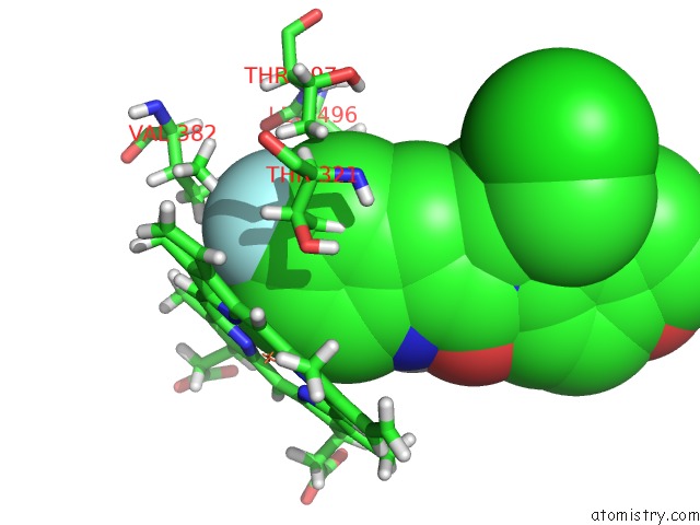
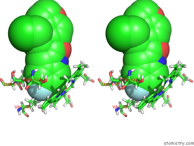
Fluorine binding site 1 out of 4 in 6udm
Go back to
Fluorine binding site 1 out
of 4 in the Structure of Human Cytochrome P450 1A1 with Duocarmycin Prodrug (S) Ict-2726
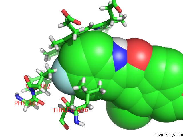
Mono view
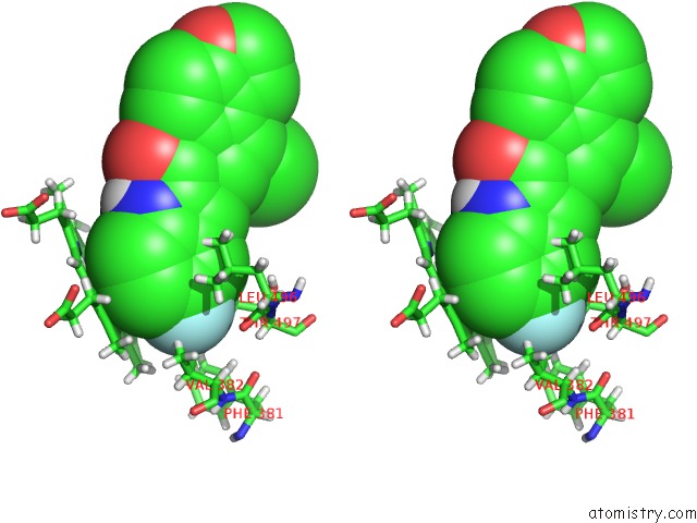
Stereo pair view
Mono view
Stereo pair view
A full contact list of Fluorine with other atoms in the F binding
site number 1 of Structure of Human Cytochrome P450 1A1 with Duocarmycin Prodrug (S) Ict-2726 within 5.0Å range:
|
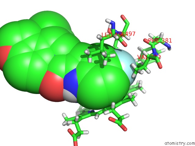
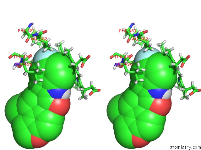
Fluorine binding site 2 out of 4 in 6udm
Go back to
Fluorine binding site 2 out
of 4 in the Structure of Human Cytochrome P450 1A1 with Duocarmycin Prodrug (S) Ict-2726
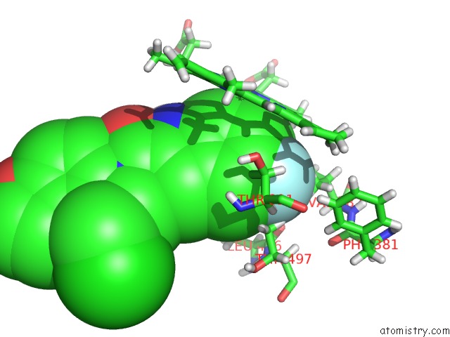
Mono view
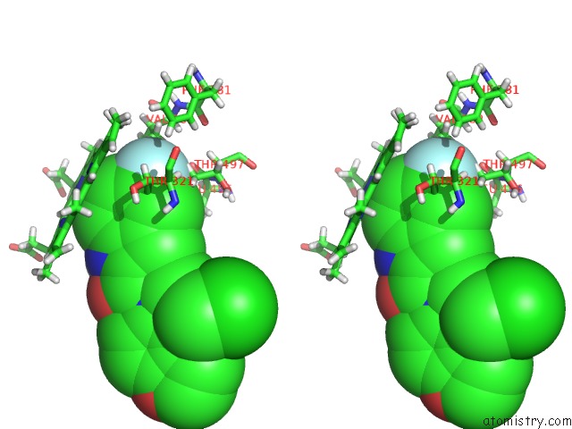
Stereo pair view
Mono view
Stereo pair view
A full contact list of Fluorine with other atoms in the F binding
site number 2 of Structure of Human Cytochrome P450 1A1 with Duocarmycin Prodrug (S) Ict-2726 within 5.0Å range:
|
Fluorine binding site 3 out of 4 in 6udm
Go back to
Fluorine binding site 3 out
of 4 in the Structure of Human Cytochrome P450 1A1 with Duocarmycin Prodrug (S) Ict-2726
Mono view
Stereo pair view
Mono view
Stereo pair view
A full contact list of Fluorine with other atoms in the F binding
site number 3 of Structure of Human Cytochrome P450 1A1 with Duocarmycin Prodrug (S) Ict-2726 within 5.0Å range:
|
Fluorine binding site 4 out of 4 in 6udm
Go back to
Fluorine binding site 4 out
of 4 in the Structure of Human Cytochrome P450 1A1 with Duocarmycin Prodrug (S) Ict-2726
Mono view
Stereo pair view
Mono view
Stereo pair view
A full contact list of Fluorine with other atoms in the F binding
site number 4 of Structure of Human Cytochrome P450 1A1 with Duocarmycin Prodrug (S) Ict-2726 within 5.0Å range:
|
Reference:
A.G.Bart,
E.E.Scott.
Designed Duocarmycin Prodrugs For Human Cytochrome P450 1A1 and 2W1 To Be Published.
Page generated: Tue Jul 15 16:15:52 2025
Last articles
Fe in 2YXOFe in 2YRS
Fe in 2YXC
Fe in 2YNM
Fe in 2YVJ
Fe in 2YP1
Fe in 2YU2
Fe in 2YU1
Fe in 2YQB
Fe in 2YOO