Fluorine »
PDB 6v3h-6vix »
6vg0 »
Fluorine in PDB 6vg0: Crystal Structure of Human Cytosolic Isocitrate Dehydrogenase (IDH1) R132H Mutant in Complex with Nadph and Agi-15056
Enzymatic activity of Crystal Structure of Human Cytosolic Isocitrate Dehydrogenase (IDH1) R132H Mutant in Complex with Nadph and Agi-15056
All present enzymatic activity of Crystal Structure of Human Cytosolic Isocitrate Dehydrogenase (IDH1) R132H Mutant in Complex with Nadph and Agi-15056:
1.1.1.42;
1.1.1.42;
Protein crystallography data
The structure of Crystal Structure of Human Cytosolic Isocitrate Dehydrogenase (IDH1) R132H Mutant in Complex with Nadph and Agi-15056, PDB code: 6vg0
was solved by
A.Padyana,
L.Jin,
with X-Ray Crystallography technique. A brief refinement statistics is given in the table below:
| Resolution Low / High (Å) | 41.20 / 2.66 |
| Space group | P 21 21 2 |
| Cell size a, b, c (Å), α, β, γ (°) | 196.770, 89.160, 90.750, 90.00, 90.00, 90.00 |
| R / Rfree (%) | 20.6 / 26.4 |
Fluorine Binding Sites:
The binding sites of Fluorine atom in the Crystal Structure of Human Cytosolic Isocitrate Dehydrogenase (IDH1) R132H Mutant in Complex with Nadph and Agi-15056
(pdb code 6vg0). This binding sites where shown within
5.0 Angstroms radius around Fluorine atom.
In total 9 binding sites of Fluorine where determined in the Crystal Structure of Human Cytosolic Isocitrate Dehydrogenase (IDH1) R132H Mutant in Complex with Nadph and Agi-15056, PDB code: 6vg0:
Jump to Fluorine binding site number: 1; 2; 3; 4; 5; 6; 7; 8; 9;
In total 9 binding sites of Fluorine where determined in the Crystal Structure of Human Cytosolic Isocitrate Dehydrogenase (IDH1) R132H Mutant in Complex with Nadph and Agi-15056, PDB code: 6vg0:
Jump to Fluorine binding site number: 1; 2; 3; 4; 5; 6; 7; 8; 9;
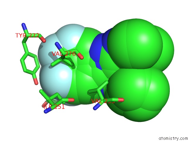
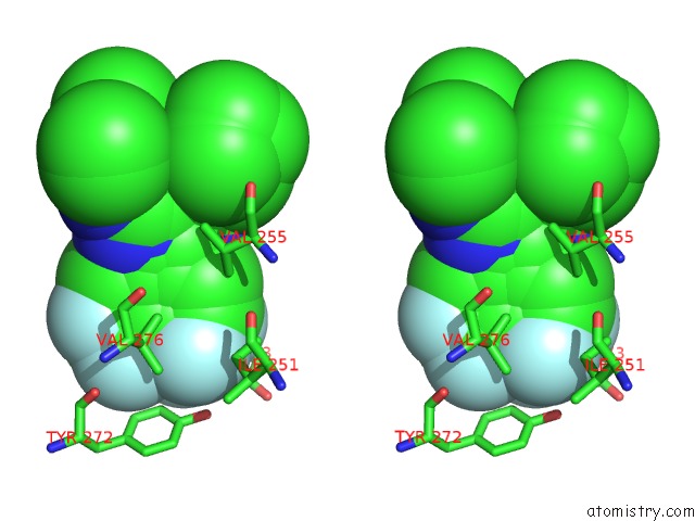
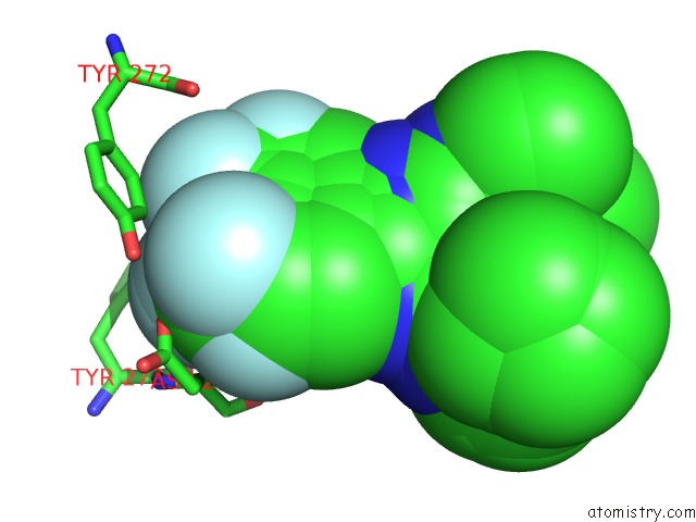
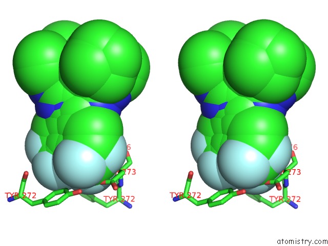
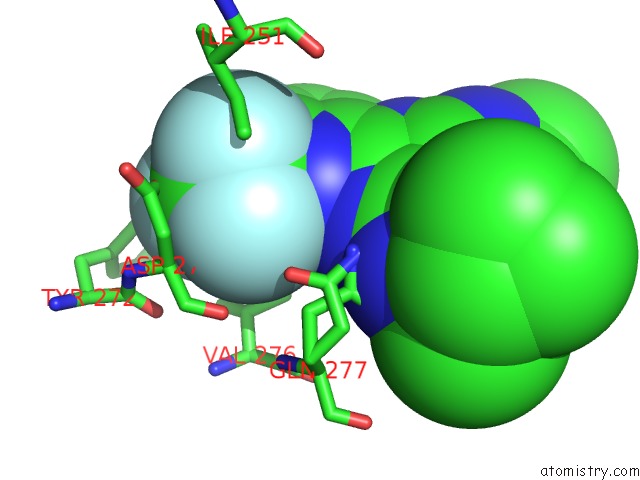
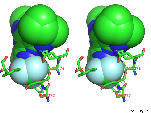
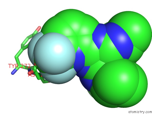
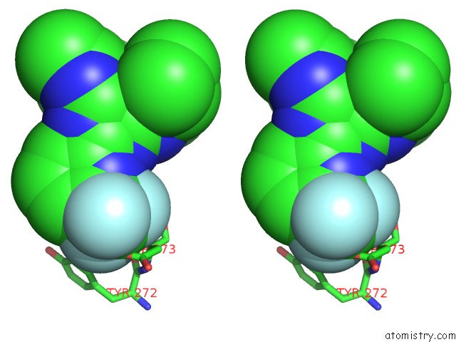
Fluorine binding site 1 out of 9 in 6vg0
Go back to
Fluorine binding site 1 out
of 9 in the Crystal Structure of Human Cytosolic Isocitrate Dehydrogenase (IDH1) R132H Mutant in Complex with Nadph and Agi-15056
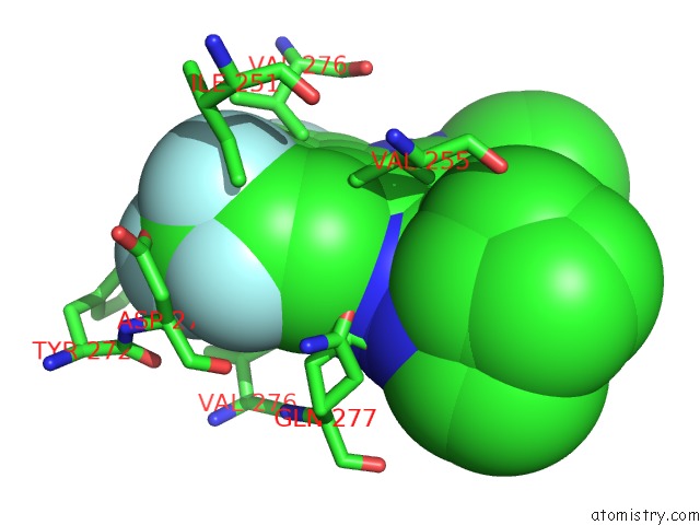
Mono view
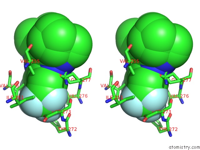
Stereo pair view
Mono view
Stereo pair view
A full contact list of Fluorine with other atoms in the F binding
site number 1 of Crystal Structure of Human Cytosolic Isocitrate Dehydrogenase (IDH1) R132H Mutant in Complex with Nadph and Agi-15056 within 5.0Å range:
|
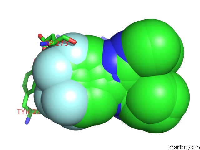
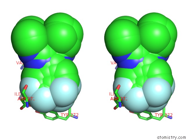
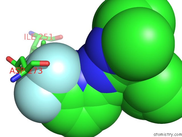
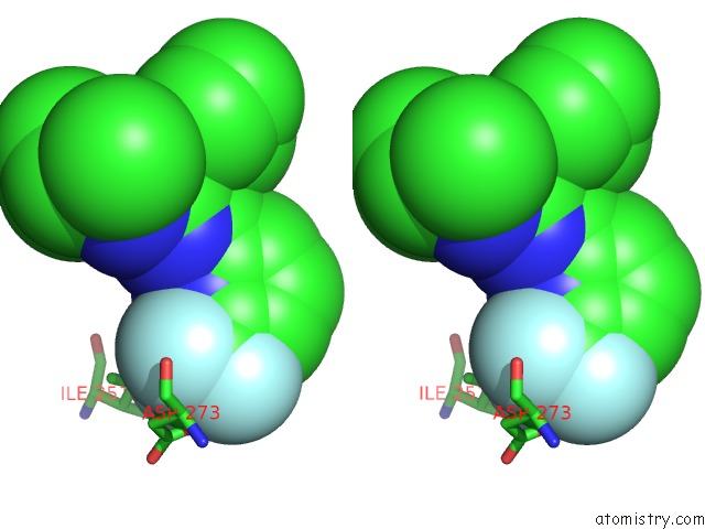
Fluorine binding site 2 out of 9 in 6vg0
Go back to
Fluorine binding site 2 out
of 9 in the Crystal Structure of Human Cytosolic Isocitrate Dehydrogenase (IDH1) R132H Mutant in Complex with Nadph and Agi-15056
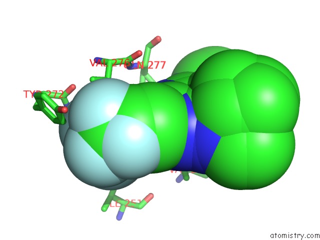
Mono view
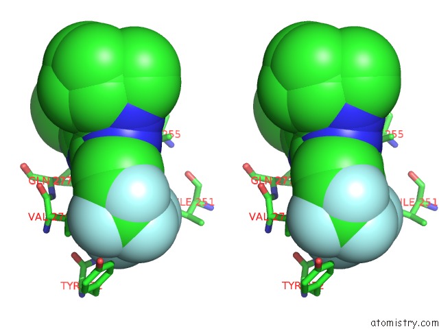
Stereo pair view
Mono view
Stereo pair view
A full contact list of Fluorine with other atoms in the F binding
site number 2 of Crystal Structure of Human Cytosolic Isocitrate Dehydrogenase (IDH1) R132H Mutant in Complex with Nadph and Agi-15056 within 5.0Å range:
|
Fluorine binding site 3 out of 9 in 6vg0
Go back to
Fluorine binding site 3 out
of 9 in the Crystal Structure of Human Cytosolic Isocitrate Dehydrogenase (IDH1) R132H Mutant in Complex with Nadph and Agi-15056
Mono view
Stereo pair view
Mono view
Stereo pair view
A full contact list of Fluorine with other atoms in the F binding
site number 3 of Crystal Structure of Human Cytosolic Isocitrate Dehydrogenase (IDH1) R132H Mutant in Complex with Nadph and Agi-15056 within 5.0Å range:
|
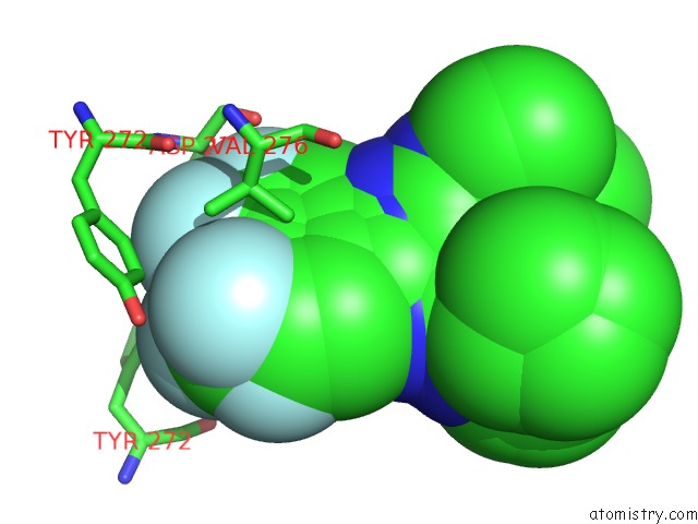
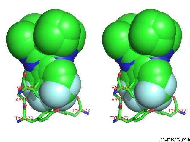
Fluorine binding site 4 out of 9 in 6vg0
Go back to
Fluorine binding site 4 out
of 9 in the Crystal Structure of Human Cytosolic Isocitrate Dehydrogenase (IDH1) R132H Mutant in Complex with Nadph and Agi-15056
Mono view
Stereo pair view
Mono view
Stereo pair view
A full contact list of Fluorine with other atoms in the F binding
site number 4 of Crystal Structure of Human Cytosolic Isocitrate Dehydrogenase (IDH1) R132H Mutant in Complex with Nadph and Agi-15056 within 5.0Å range:
|
Fluorine binding site 5 out of 9 in 6vg0
Go back to
Fluorine binding site 5 out
of 9 in the Crystal Structure of Human Cytosolic Isocitrate Dehydrogenase (IDH1) R132H Mutant in Complex with Nadph and Agi-15056
Mono view
Stereo pair view
Mono view
Stereo pair view
A full contact list of Fluorine with other atoms in the F binding
site number 5 of Crystal Structure of Human Cytosolic Isocitrate Dehydrogenase (IDH1) R132H Mutant in Complex with Nadph and Agi-15056 within 5.0Å range:
|
Fluorine binding site 6 out of 9 in 6vg0
Go back to
Fluorine binding site 6 out
of 9 in the Crystal Structure of Human Cytosolic Isocitrate Dehydrogenase (IDH1) R132H Mutant in Complex with Nadph and Agi-15056
Mono view
Stereo pair view
Mono view
Stereo pair view
A full contact list of Fluorine with other atoms in the F binding
site number 6 of Crystal Structure of Human Cytosolic Isocitrate Dehydrogenase (IDH1) R132H Mutant in Complex with Nadph and Agi-15056 within 5.0Å range:
|
Fluorine binding site 7 out of 9 in 6vg0
Go back to
Fluorine binding site 7 out
of 9 in the Crystal Structure of Human Cytosolic Isocitrate Dehydrogenase (IDH1) R132H Mutant in Complex with Nadph and Agi-15056
Mono view
Stereo pair view
Mono view
Stereo pair view
A full contact list of Fluorine with other atoms in the F binding
site number 7 of Crystal Structure of Human Cytosolic Isocitrate Dehydrogenase (IDH1) R132H Mutant in Complex with Nadph and Agi-15056 within 5.0Å range:
|
Fluorine binding site 8 out of 9 in 6vg0
Go back to
Fluorine binding site 8 out
of 9 in the Crystal Structure of Human Cytosolic Isocitrate Dehydrogenase (IDH1) R132H Mutant in Complex with Nadph and Agi-15056
Mono view
Stereo pair view
Mono view
Stereo pair view
A full contact list of Fluorine with other atoms in the F binding
site number 8 of Crystal Structure of Human Cytosolic Isocitrate Dehydrogenase (IDH1) R132H Mutant in Complex with Nadph and Agi-15056 within 5.0Å range:
|
Fluorine binding site 9 out of 9 in 6vg0
Go back to
Fluorine binding site 9 out
of 9 in the Crystal Structure of Human Cytosolic Isocitrate Dehydrogenase (IDH1) R132H Mutant in Complex with Nadph and Agi-15056
Mono view
Stereo pair view
Mono view
Stereo pair view
A full contact list of Fluorine with other atoms in the F binding
site number 9 of Crystal Structure of Human Cytosolic Isocitrate Dehydrogenase (IDH1) R132H Mutant in Complex with Nadph and Agi-15056 within 5.0Å range:
|
Reference:
Z.Konteatis,
E.Artin,
B.Nicolay,
K.Straley,
A.K.Padyana,
L.Jin,
Y.Chen,
R.Narayaraswamy,
S.Tong,
F.Wang,
D.Zhou,
D.Cui,
Z.Cai,
Z.Luo,
C.Fang,
H.Tang,
X.Lv,
R.Nagaraja,
H.Yang,
S.M.Su,
Z.Sui,
L.Dang,
K.Yen,
J.Popovici-Muller,
P.Codega,
C.Campos,
I.K.Mellinghoff,
S.Biller.
Vorasidenib (Ag-881): A First-in-Class, Brain-Penetrant Dual Inhibitor of Mutant IDH1 and 2 For Treatment of Glioma Acs Med.Chem.Lett. 2020.
ISSN: ISSN 1948-5875
DOI: 10.1021/ACSMEDCHEMLETT.9B00509
Page generated: Tue Jul 15 16:38:28 2025
ISSN: ISSN 1948-5875
DOI: 10.1021/ACSMEDCHEMLETT.9B00509
Last articles
Fe in 2YXOFe in 2YRS
Fe in 2YXC
Fe in 2YNM
Fe in 2YVJ
Fe in 2YP1
Fe in 2YU2
Fe in 2YU1
Fe in 2YQB
Fe in 2YOO