Fluorine »
PDB 6x3c-6xk0 »
6x3c »
Fluorine in PDB 6x3c: Crystal Structure of Streptogramin A Acetyltransferase Vata From Staphylococcus Aureus in Complex with Streptogramin Analog F1037 (47)
Protein crystallography data
The structure of Crystal Structure of Streptogramin A Acetyltransferase Vata From Staphylococcus Aureus in Complex with Streptogramin Analog F1037 (47), PDB code: 6x3c
was solved by
H.A.Chaires,
J.S.Fraser,
with X-Ray Crystallography technique. A brief refinement statistics is given in the table below:
| Resolution Low / High (Å) | 89.14 / 3.05 |
| Space group | P 21 21 21 |
| Cell size a, b, c (Å), α, β, γ (°) | 105.540, 105.620, 178.200, 90.00, 90.00, 90.00 |
| R / Rfree (%) | 23.4 / 26.6 |
Other elements in 6x3c:
The structure of Crystal Structure of Streptogramin A Acetyltransferase Vata From Staphylococcus Aureus in Complex with Streptogramin Analog F1037 (47) also contains other interesting chemical elements:
| Magnesium | (Mg) | 2 atoms |
| Chlorine | (Cl) | 6 atoms |
Fluorine Binding Sites:
The binding sites of Fluorine atom in the Crystal Structure of Streptogramin A Acetyltransferase Vata From Staphylococcus Aureus in Complex with Streptogramin Analog F1037 (47)
(pdb code 6x3c). This binding sites where shown within
5.0 Angstroms radius around Fluorine atom.
In total 6 binding sites of Fluorine where determined in the Crystal Structure of Streptogramin A Acetyltransferase Vata From Staphylococcus Aureus in Complex with Streptogramin Analog F1037 (47), PDB code: 6x3c:
Jump to Fluorine binding site number: 1; 2; 3; 4; 5; 6;
In total 6 binding sites of Fluorine where determined in the Crystal Structure of Streptogramin A Acetyltransferase Vata From Staphylococcus Aureus in Complex with Streptogramin Analog F1037 (47), PDB code: 6x3c:
Jump to Fluorine binding site number: 1; 2; 3; 4; 5; 6;
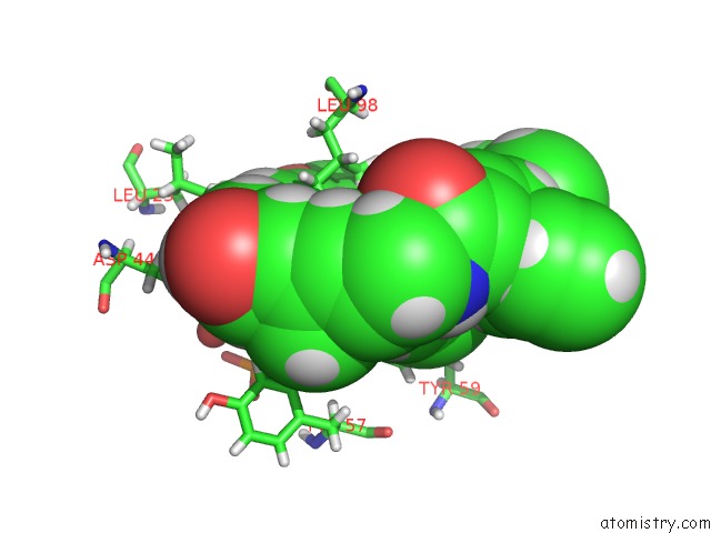
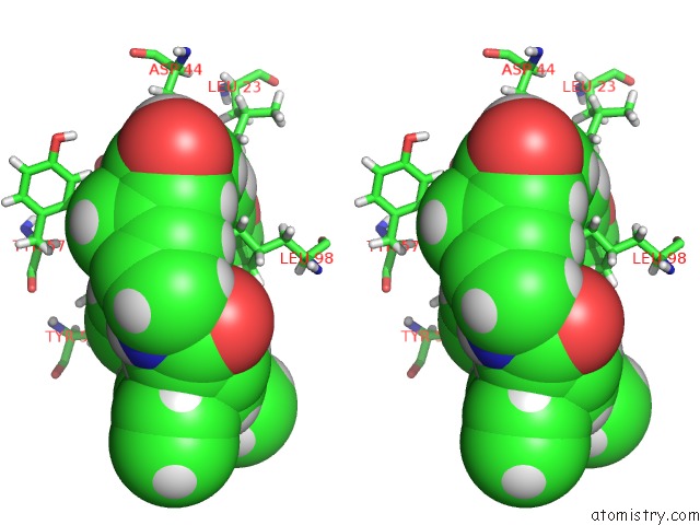
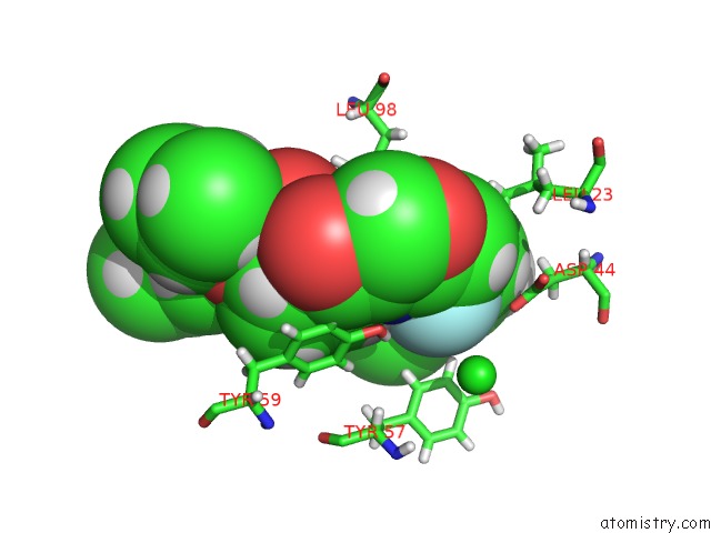
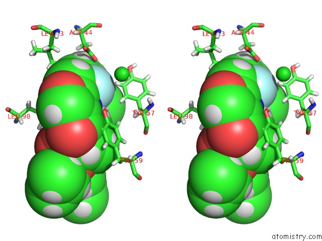
Fluorine binding site 1 out of 6 in 6x3c
Go back to
Fluorine binding site 1 out
of 6 in the Crystal Structure of Streptogramin A Acetyltransferase Vata From Staphylococcus Aureus in Complex with Streptogramin Analog F1037 (47)
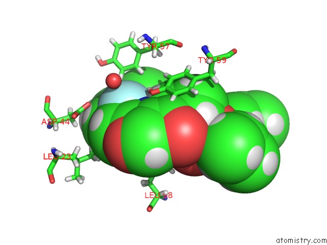
Mono view

Stereo pair view
Mono view
Stereo pair view
A full contact list of Fluorine with other atoms in the F binding
site number 1 of Crystal Structure of Streptogramin A Acetyltransferase Vata From Staphylococcus Aureus in Complex with Streptogramin Analog F1037 (47) within 5.0Å range:
|
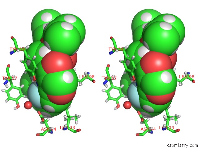
Fluorine binding site 2 out of 6 in 6x3c
Go back to
Fluorine binding site 2 out
of 6 in the Crystal Structure of Streptogramin A Acetyltransferase Vata From Staphylococcus Aureus in Complex with Streptogramin Analog F1037 (47)
Mono view

Stereo pair view
Mono view
Stereo pair view
A full contact list of Fluorine with other atoms in the F binding
site number 2 of Crystal Structure of Streptogramin A Acetyltransferase Vata From Staphylococcus Aureus in Complex with Streptogramin Analog F1037 (47) within 5.0Å range:
|
Fluorine binding site 3 out of 6 in 6x3c
Go back to
Fluorine binding site 3 out
of 6 in the Crystal Structure of Streptogramin A Acetyltransferase Vata From Staphylococcus Aureus in Complex with Streptogramin Analog F1037 (47)

Mono view
Stereo pair view
Mono view
Stereo pair view
A full contact list of Fluorine with other atoms in the F binding
site number 3 of Crystal Structure of Streptogramin A Acetyltransferase Vata From Staphylococcus Aureus in Complex with Streptogramin Analog F1037 (47) within 5.0Å range:
|
Fluorine binding site 4 out of 6 in 6x3c
Go back to
Fluorine binding site 4 out
of 6 in the Crystal Structure of Streptogramin A Acetyltransferase Vata From Staphylococcus Aureus in Complex with Streptogramin Analog F1037 (47)

Mono view

Stereo pair view
Mono view
Stereo pair view
A full contact list of Fluorine with other atoms in the F binding
site number 4 of Crystal Structure of Streptogramin A Acetyltransferase Vata From Staphylococcus Aureus in Complex with Streptogramin Analog F1037 (47) within 5.0Å range:
|
Fluorine binding site 5 out of 6 in 6x3c
Go back to
Fluorine binding site 5 out
of 6 in the Crystal Structure of Streptogramin A Acetyltransferase Vata From Staphylococcus Aureus in Complex with Streptogramin Analog F1037 (47)
Mono view
Stereo pair view
Mono view
Stereo pair view
A full contact list of Fluorine with other atoms in the F binding
site number 5 of Crystal Structure of Streptogramin A Acetyltransferase Vata From Staphylococcus Aureus in Complex with Streptogramin Analog F1037 (47) within 5.0Å range:
|
Fluorine binding site 6 out of 6 in 6x3c
Go back to
Fluorine binding site 6 out
of 6 in the Crystal Structure of Streptogramin A Acetyltransferase Vata From Staphylococcus Aureus in Complex with Streptogramin Analog F1037 (47)

Mono view
Stereo pair view
Mono view
Stereo pair view
A full contact list of Fluorine with other atoms in the F binding
site number 6 of Crystal Structure of Streptogramin A Acetyltransferase Vata From Staphylococcus Aureus in Complex with Streptogramin Analog F1037 (47) within 5.0Å range:
|
Reference:
Q.Li,
J.Pellegrino,
D.J.Lee,
A.A.Tran,
H.A.Chaires,
R.Wang,
J.E.Park,
K.Ji,
D.Chow,
N.Zhang,
A.F.Brilot,
J.T.Biel,
G.Van Zundert,
K.Borrelli,
D.Shinabarger,
C.Wolfe,
B.Murray,
M.P.Jacobson,
E.Muhle,
O.Chesneau,
J.S.Fraser,
I.B.Seiple.
Synthetic Group A Streptogramin Antibiotics That Overcome Vat Resistance. Nature V. 586 145 2020.
ISSN: ESSN 1476-4687
PubMed: 32968273
DOI: 10.1038/S41586-020-2761-3
Page generated: Fri Aug 2 03:59:28 2024
ISSN: ESSN 1476-4687
PubMed: 32968273
DOI: 10.1038/S41586-020-2761-3
Last articles
Zn in 9J0NZn in 9J0O
Zn in 9J0P
Zn in 9FJX
Zn in 9EKB
Zn in 9C0F
Zn in 9CAH
Zn in 9CH0
Zn in 9CH3
Zn in 9CH1