Fluorine »
PDB 7aym-7biv »
7b33 »
Fluorine in PDB 7b33: MST3 in Complex with MRIA11
Enzymatic activity of MST3 in Complex with MRIA11
All present enzymatic activity of MST3 in Complex with MRIA11:
2.7.11.1;
2.7.11.1;
Protein crystallography data
The structure of MST3 in Complex with MRIA11, PDB code: 7b33
was solved by
R.Tesch,
M.Rak,
A.C.Joerger,
S.Knapp,
Structural Genomics Consortium(Sgc),
with X-Ray Crystallography technique. A brief refinement statistics is given in the table below:
| Resolution Low / High (Å) | 49.41 / 1.90 |
| Space group | C 1 2 1 |
| Cell size a, b, c (Å), α, β, γ (°) | 98.972, 58.954, 61.441, 90.00, 93.13, 90.00 |
| R / Rfree (%) | 18.1 / 20.8 |
Other elements in 7b33:
The structure of MST3 in Complex with MRIA11 also contains other interesting chemical elements:
| Chlorine | (Cl) | 1 atom |
Fluorine Binding Sites:
The binding sites of Fluorine atom in the MST3 in Complex with MRIA11
(pdb code 7b33). This binding sites where shown within
5.0 Angstroms radius around Fluorine atom.
In total 2 binding sites of Fluorine where determined in the MST3 in Complex with MRIA11, PDB code: 7b33:
Jump to Fluorine binding site number: 1; 2;
In total 2 binding sites of Fluorine where determined in the MST3 in Complex with MRIA11, PDB code: 7b33:
Jump to Fluorine binding site number: 1; 2;
Fluorine binding site 1 out of 2 in 7b33
Go back to
Fluorine binding site 1 out
of 2 in the MST3 in Complex with MRIA11
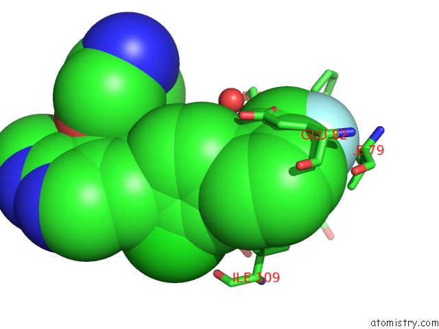
Mono view
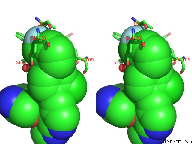
Stereo pair view
Mono view
Stereo pair view
A full contact list of Fluorine with other atoms in the F binding
site number 1 of MST3 in Complex with MRIA11 within 5.0Å range:
|
Fluorine binding site 2 out of 2 in 7b33
Go back to
Fluorine binding site 2 out
of 2 in the MST3 in Complex with MRIA11
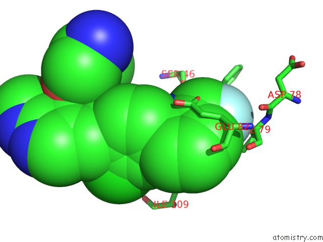
Mono view
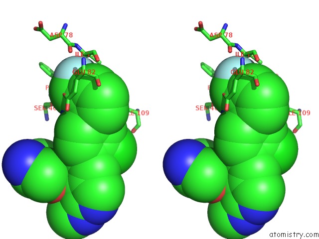
Stereo pair view
Mono view
Stereo pair view
A full contact list of Fluorine with other atoms in the F binding
site number 2 of MST3 in Complex with MRIA11 within 5.0Å range:
|
Reference:
R.Tesch,
M.Rak,
A.C.Joerger,
S.Knapp,
Structural Genomics Consortium (Sgc).
Structure-Based Design of Selective Salt-Inducible Kinase (Sik) Inhibitors To Be Published.
Page generated: Fri Aug 2 05:54:54 2024
Last articles
Zn in 9JYWZn in 9IR4
Zn in 9IR3
Zn in 9GMX
Zn in 9GMW
Zn in 9JEJ
Zn in 9ERF
Zn in 9ERE
Zn in 9EGV
Zn in 9EGW