Fluorine »
PDB 7az1-7bj6 »
7b9o »
Fluorine in PDB 7b9o: Crystal Structure of Retinoic Acid Receptor Alpha (Rxra) in Complexed with S169 Inhibitor
Protein crystallography data
The structure of Crystal Structure of Retinoic Acid Receptor Alpha (Rxra) in Complexed with S169 Inhibitor, PDB code: 7b9o
was solved by
X.Ni,
A.Chaikuad,
S.Schierle,
D.Merk,
S.Knapp,
Structural Genomicsconsortium (Sgc),
with X-Ray Crystallography technique. A brief refinement statistics is given in the table below:
| Resolution Low / High (Å) | 46.34 / 2.05 |
| Space group | P 43 21 2 |
| Cell size a, b, c (Å), α, β, γ (°) | 65.529, 65.529, 110.178, 90, 90, 90 |
| R / Rfree (%) | 19.7 / 23.6 |
Fluorine Binding Sites:
The binding sites of Fluorine atom in the Crystal Structure of Retinoic Acid Receptor Alpha (Rxra) in Complexed with S169 Inhibitor
(pdb code 7b9o). This binding sites where shown within
5.0 Angstroms radius around Fluorine atom.
In total 6 binding sites of Fluorine where determined in the Crystal Structure of Retinoic Acid Receptor Alpha (Rxra) in Complexed with S169 Inhibitor, PDB code: 7b9o:
Jump to Fluorine binding site number: 1; 2; 3; 4; 5; 6;
In total 6 binding sites of Fluorine where determined in the Crystal Structure of Retinoic Acid Receptor Alpha (Rxra) in Complexed with S169 Inhibitor, PDB code: 7b9o:
Jump to Fluorine binding site number: 1; 2; 3; 4; 5; 6;
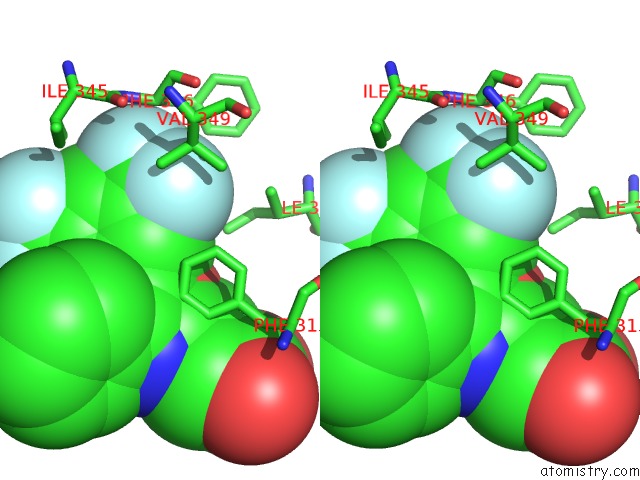
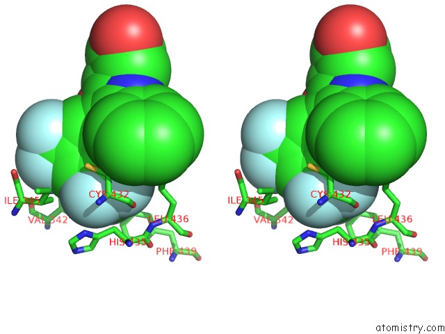
Fluorine binding site 1 out of 6 in 7b9o
Go back to
Fluorine binding site 1 out
of 6 in the Crystal Structure of Retinoic Acid Receptor Alpha (Rxra) in Complexed with S169 Inhibitor
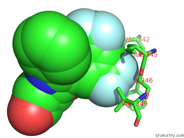
Mono view
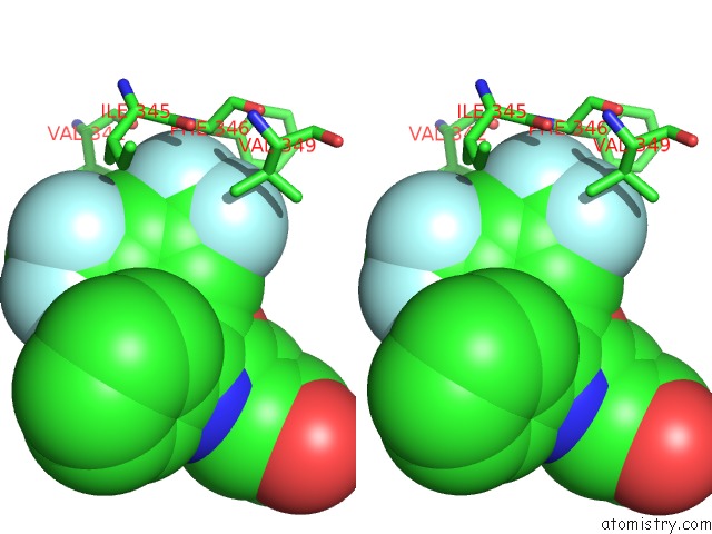
Stereo pair view
Mono view
Stereo pair view
A full contact list of Fluorine with other atoms in the F binding
site number 1 of Crystal Structure of Retinoic Acid Receptor Alpha (Rxra) in Complexed with S169 Inhibitor within 5.0Å range:
|
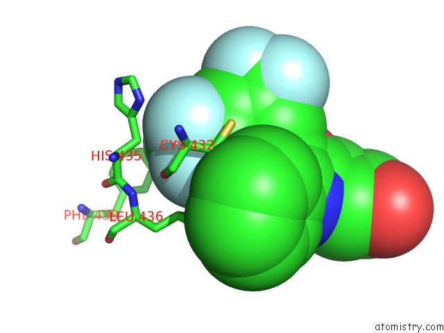
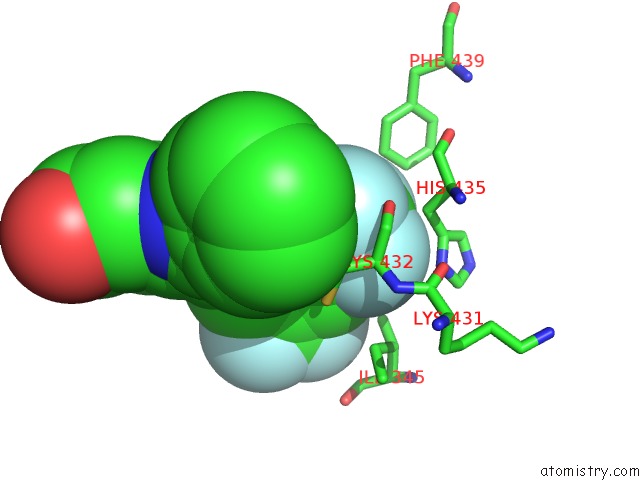
Fluorine binding site 2 out of 6 in 7b9o
Go back to
Fluorine binding site 2 out
of 6 in the Crystal Structure of Retinoic Acid Receptor Alpha (Rxra) in Complexed with S169 Inhibitor

Mono view
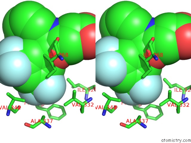
Stereo pair view
Mono view
Stereo pair view
A full contact list of Fluorine with other atoms in the F binding
site number 2 of Crystal Structure of Retinoic Acid Receptor Alpha (Rxra) in Complexed with S169 Inhibitor within 5.0Å range:
|
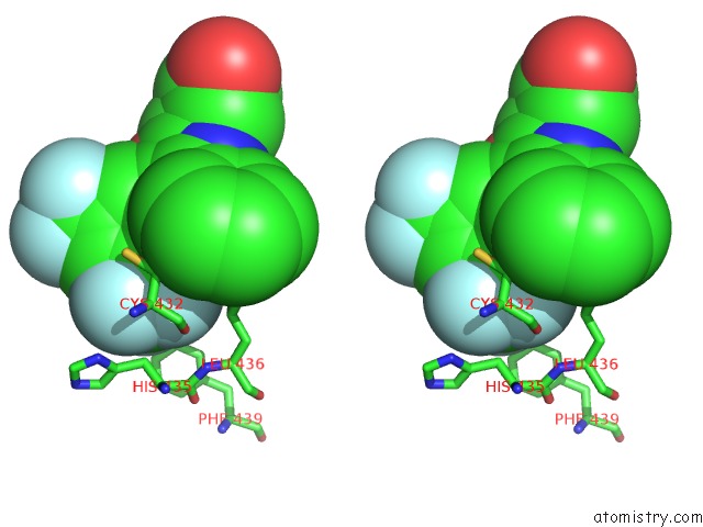
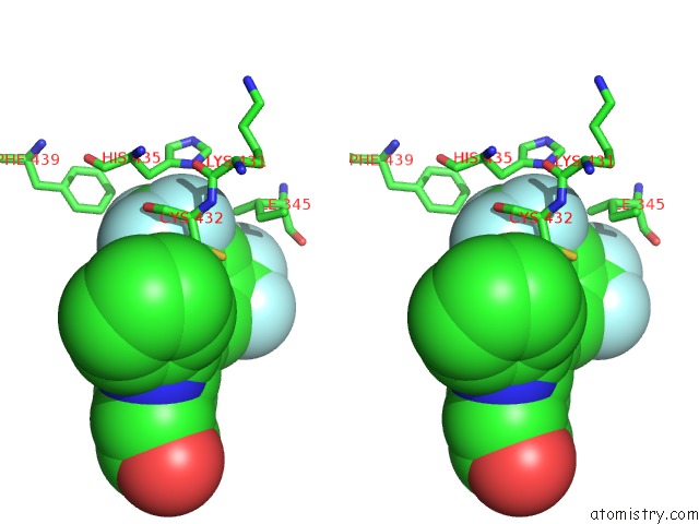
Fluorine binding site 3 out of 6 in 7b9o
Go back to
Fluorine binding site 3 out
of 6 in the Crystal Structure of Retinoic Acid Receptor Alpha (Rxra) in Complexed with S169 Inhibitor
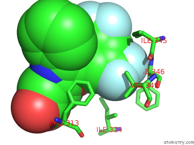
Mono view
Stereo pair view
Mono view
Stereo pair view
A full contact list of Fluorine with other atoms in the F binding
site number 3 of Crystal Structure of Retinoic Acid Receptor Alpha (Rxra) in Complexed with S169 Inhibitor within 5.0Å range:
|
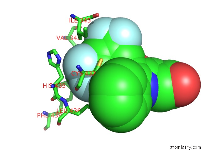
Fluorine binding site 4 out of 6 in 7b9o
Go back to
Fluorine binding site 4 out
of 6 in the Crystal Structure of Retinoic Acid Receptor Alpha (Rxra) in Complexed with S169 Inhibitor
Mono view
Stereo pair view
Mono view
Stereo pair view
A full contact list of Fluorine with other atoms in the F binding
site number 4 of Crystal Structure of Retinoic Acid Receptor Alpha (Rxra) in Complexed with S169 Inhibitor within 5.0Å range:
|
Fluorine binding site 5 out of 6 in 7b9o
Go back to
Fluorine binding site 5 out
of 6 in the Crystal Structure of Retinoic Acid Receptor Alpha (Rxra) in Complexed with S169 Inhibitor
Mono view
Stereo pair view
Mono view
Stereo pair view
A full contact list of Fluorine with other atoms in the F binding
site number 5 of Crystal Structure of Retinoic Acid Receptor Alpha (Rxra) in Complexed with S169 Inhibitor within 5.0Å range:
|
Fluorine binding site 6 out of 6 in 7b9o
Go back to
Fluorine binding site 6 out
of 6 in the Crystal Structure of Retinoic Acid Receptor Alpha (Rxra) in Complexed with S169 Inhibitor
Mono view
Stereo pair view
Mono view
Stereo pair view
A full contact list of Fluorine with other atoms in the F binding
site number 6 of Crystal Structure of Retinoic Acid Receptor Alpha (Rxra) in Complexed with S169 Inhibitor within 5.0Å range:
|
Reference:
X.Ni,
A.Chaikuad,
S.Schierle,
D.Merk,
S.Knapp,
Structural Genomics Consortium (Sgc).
Crystal Structure of Retinoic Acid Receptor Alpha (Rxra) in Complexed with S169 Inhibitor To Be Published.
Page generated: Tue Jul 15 18:45:34 2025
Last articles
Fe in 2YXOFe in 2YRS
Fe in 2YXC
Fe in 2YNM
Fe in 2YVJ
Fe in 2YP1
Fe in 2YU2
Fe in 2YU1
Fe in 2YQB
Fe in 2YOO