Fluorine »
PDB 7cky-7db4 »
7cwf »
Fluorine in PDB 7cwf: Crystal Structure of PDE8A Catalytic Domain in Complex with 2C
Enzymatic activity of Crystal Structure of PDE8A Catalytic Domain in Complex with 2C
All present enzymatic activity of Crystal Structure of PDE8A Catalytic Domain in Complex with 2C:
3.1.4.53;
3.1.4.53;
Protein crystallography data
The structure of Crystal Structure of PDE8A Catalytic Domain in Complex with 2C, PDB code: 7cwf
was solved by
Y.Huang,
X.-N.Wu,
Q.Zhou,
Y.Wu,
H.-B.Luo,
with X-Ray Crystallography technique. A brief refinement statistics is given in the table below:
| Resolution Low / High (Å) | 23.66 / 2.80 |
| Space group | C 2 2 21 |
| Cell size a, b, c (Å), α, β, γ (°) | 76.114, 131.874, 101.398, 90, 90, 90 |
| R / Rfree (%) | 25.7 / 29.3 |
Other elements in 7cwf:
The structure of Crystal Structure of PDE8A Catalytic Domain in Complex with 2C also contains other interesting chemical elements:
| Zinc | (Zn) | 1 atom |
| Magnesium | (Mg) | 1 atom |
| Chlorine | (Cl) | 1 atom |
Fluorine Binding Sites:
The binding sites of Fluorine atom in the Crystal Structure of PDE8A Catalytic Domain in Complex with 2C
(pdb code 7cwf). This binding sites where shown within
5.0 Angstroms radius around Fluorine atom.
In total 2 binding sites of Fluorine where determined in the Crystal Structure of PDE8A Catalytic Domain in Complex with 2C, PDB code: 7cwf:
Jump to Fluorine binding site number: 1; 2;
In total 2 binding sites of Fluorine where determined in the Crystal Structure of PDE8A Catalytic Domain in Complex with 2C, PDB code: 7cwf:
Jump to Fluorine binding site number: 1; 2;
Fluorine binding site 1 out of 2 in 7cwf
Go back to
Fluorine binding site 1 out
of 2 in the Crystal Structure of PDE8A Catalytic Domain in Complex with 2C
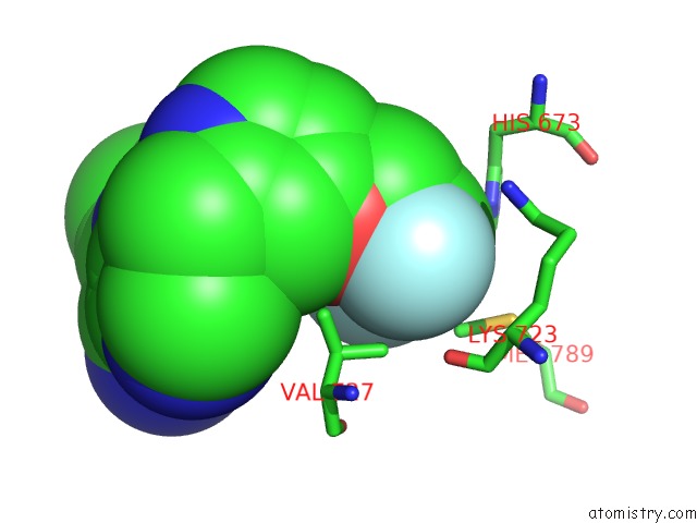
Mono view
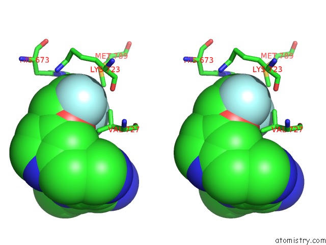
Stereo pair view
Mono view
Stereo pair view
A full contact list of Fluorine with other atoms in the F binding
site number 1 of Crystal Structure of PDE8A Catalytic Domain in Complex with 2C within 5.0Å range:
|
Fluorine binding site 2 out of 2 in 7cwf
Go back to
Fluorine binding site 2 out
of 2 in the Crystal Structure of PDE8A Catalytic Domain in Complex with 2C
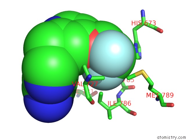
Mono view
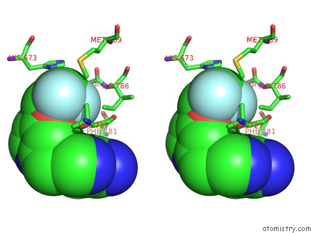
Stereo pair view
Mono view
Stereo pair view
A full contact list of Fluorine with other atoms in the F binding
site number 2 of Crystal Structure of PDE8A Catalytic Domain in Complex with 2C within 5.0Å range:
|
Reference:
Y.Huang,
X.-N.Wu,
Q.Zhou,
Y.Wu,
H.-B.Luo.
Discovery of Highly Selective and Orally Bioavailable Phosphodiesterase-8A Inhibitors. Structure-Activity Relationship, Crystal Structures, and in Vivo Studies To Be Published.
Page generated: Fri Aug 2 06:16:43 2024
Last articles
Cl in 5UFECl in 5UEU
Cl in 5UF1
Cl in 5UE1
Cl in 5UEI
Cl in 5UES
Cl in 5UEB
Cl in 5UEH
Cl in 5UE8
Cl in 5UE9