Fluorine »
PDB 7cky-7db4 »
7d9o »
Fluorine in PDB 7d9o: Crystal Structure of Recombinant Human Acetylcholinesterase in Complex with Compound 2
Enzymatic activity of Crystal Structure of Recombinant Human Acetylcholinesterase in Complex with Compound 2
All present enzymatic activity of Crystal Structure of Recombinant Human Acetylcholinesterase in Complex with Compound 2:
3.1.1.7;
3.1.1.7;
Protein crystallography data
The structure of Crystal Structure of Recombinant Human Acetylcholinesterase in Complex with Compound 2, PDB code: 7d9o
was solved by
Q.F.Liu,
W.C.Yin,
with X-Ray Crystallography technique. A brief refinement statistics is given in the table below:
| Resolution Low / High (Å) | 49.89 / 2.45 |
| Space group | P 31 2 1 |
| Cell size a, b, c (Å), α, β, γ (°) | 104.866, 104.866, 324.416, 90, 90, 120 |
| R / Rfree (%) | 18.7 / 21.4 |
Fluorine Binding Sites:
The binding sites of Fluorine atom in the Crystal Structure of Recombinant Human Acetylcholinesterase in Complex with Compound 2
(pdb code 7d9o). This binding sites where shown within
5.0 Angstroms radius around Fluorine atom.
In total 4 binding sites of Fluorine where determined in the Crystal Structure of Recombinant Human Acetylcholinesterase in Complex with Compound 2, PDB code: 7d9o:
Jump to Fluorine binding site number: 1; 2; 3; 4;
In total 4 binding sites of Fluorine where determined in the Crystal Structure of Recombinant Human Acetylcholinesterase in Complex with Compound 2, PDB code: 7d9o:
Jump to Fluorine binding site number: 1; 2; 3; 4;
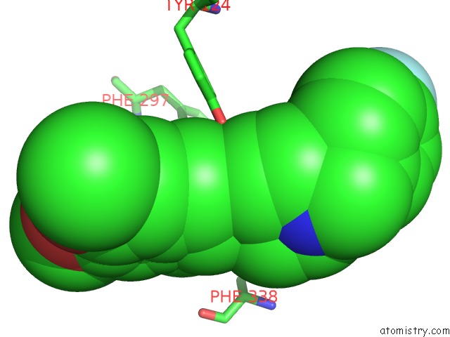
Fluorine binding site 1 out of 4 in 7d9o
Go back to
Fluorine binding site 1 out
of 4 in the Crystal Structure of Recombinant Human Acetylcholinesterase in Complex with Compound 2
Mono view

Stereo pair view
Mono view
Stereo pair view
A full contact list of Fluorine with other atoms in the F binding
site number 1 of Crystal Structure of Recombinant Human Acetylcholinesterase in Complex with Compound 2 within 5.0Å range:
|
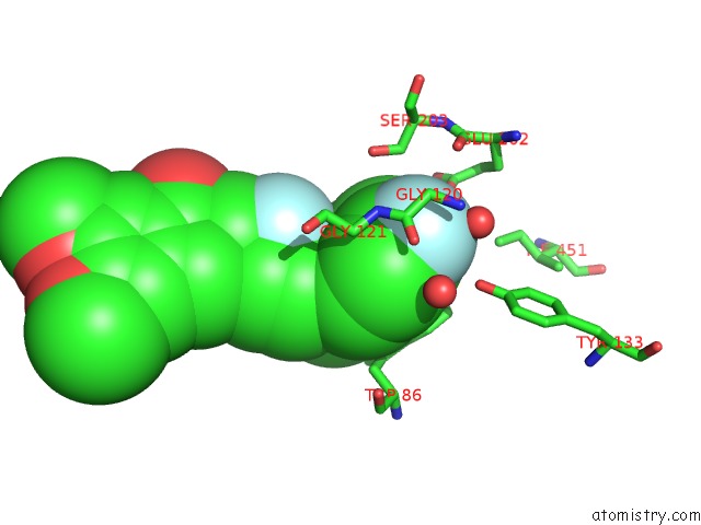
Fluorine binding site 2 out of 4 in 7d9o
Go back to
Fluorine binding site 2 out
of 4 in the Crystal Structure of Recombinant Human Acetylcholinesterase in Complex with Compound 2
Mono view

Stereo pair view
Mono view
Stereo pair view
A full contact list of Fluorine with other atoms in the F binding
site number 2 of Crystal Structure of Recombinant Human Acetylcholinesterase in Complex with Compound 2 within 5.0Å range:
|
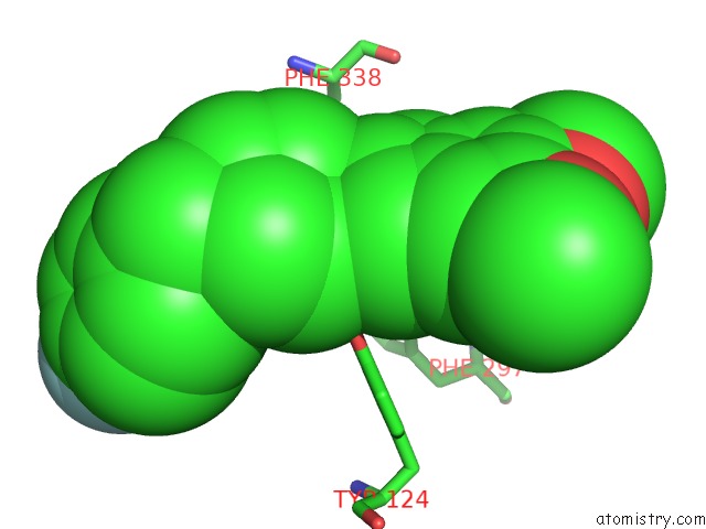
Fluorine binding site 3 out of 4 in 7d9o
Go back to
Fluorine binding site 3 out
of 4 in the Crystal Structure of Recombinant Human Acetylcholinesterase in Complex with Compound 2
Mono view

Stereo pair view
Mono view
Stereo pair view
A full contact list of Fluorine with other atoms in the F binding
site number 3 of Crystal Structure of Recombinant Human Acetylcholinesterase in Complex with Compound 2 within 5.0Å range:
|
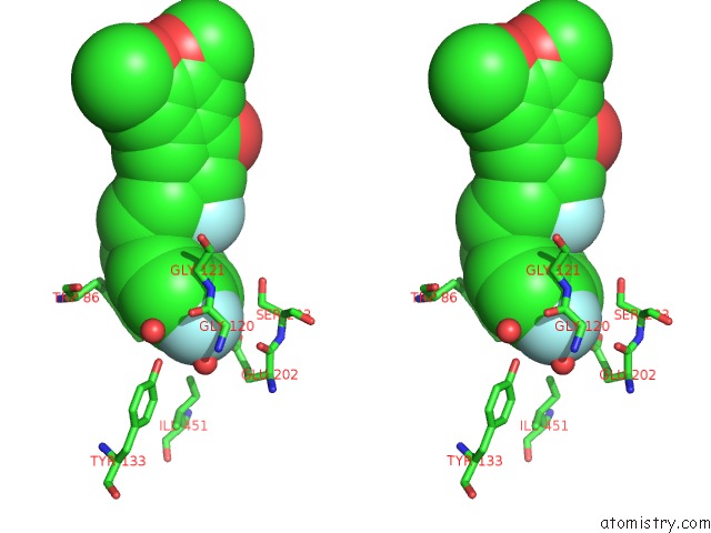
Fluorine binding site 4 out of 4 in 7d9o
Go back to
Fluorine binding site 4 out
of 4 in the Crystal Structure of Recombinant Human Acetylcholinesterase in Complex with Compound 2

Mono view
Stereo pair view
Mono view
Stereo pair view
A full contact list of Fluorine with other atoms in the F binding
site number 4 of Crystal Structure of Recombinant Human Acetylcholinesterase in Complex with Compound 2 within 5.0Å range:
|
Reference:
Y.Zhou,
Y.Fu,
W.Yin,
J.Li,
W.Wang,
F.Bai,
S.Xu,
Q.Gong,
T.Peng,
Y.Hong,
D.Zhang,
D.Zhang,
Q.Liu,
Y.Xu,
H.E.Xu,
H.Zhang,
H.Jiang,
H.Liu.
Kinetics-Driven Drug Design Strategy For Next-Generation Acetylcholinesterase Inhibitors to Clinical Candidate. J.Med.Chem. V. 64 1844 2021.
ISSN: ISSN 0022-2623
PubMed: 33570950
DOI: 10.1021/ACS.JMEDCHEM.0C01863
Page generated: Fri Aug 2 06:23:17 2024
ISSN: ISSN 0022-2623
PubMed: 33570950
DOI: 10.1021/ACS.JMEDCHEM.0C01863
Last articles
Zn in 9J0NZn in 9J0O
Zn in 9J0P
Zn in 9FJX
Zn in 9EKB
Zn in 9C0F
Zn in 9CAH
Zn in 9CH0
Zn in 9CH3
Zn in 9CH1